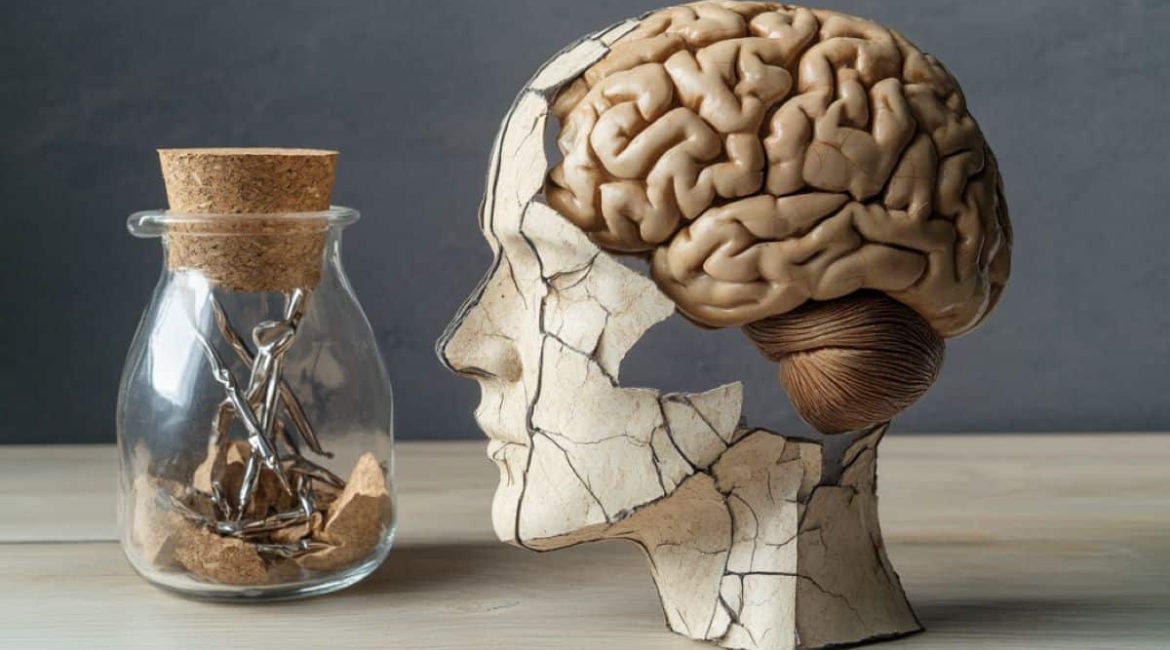
Summary: New analysis uncovers how mitophagy, the mobile recycling of damaged cells, changes automatically in the aging mammal brain. Mitophagy increases in memory-related tissue in the middle of life but quickly declines as you get older, while mitophagy increases in motor-related brain cells as you get older.
The investigation also highlights that aging cells lose acidity, mirroring alterations seen in Alzheimer’s disease designs. These findings challenge conventional wisdom regarding the universal decline of mitophagy as people age and designate the middle ages as the crucial time for neurodegenerative disease treatment.
Important Facts:
- As people get older, the interactions of mitochondria vary depending on the types of brain cells.
- Aging cells lose acidity, linking standard aging to Alzheimer ‘s-like changes.
- The middle of life is crucial to maintaining mental health.
Origin: University of Helsinki
Mitochondria, the powerhouses of our tissues, play an important part in maintaining cellular health. When damaged, they are removed through a recycling procedure called mitophagy, which is essential for the work of long-lived tissue, especially in the mind.
Impaired mitophagy has been strongly associated with neurological disorders like Alzheimer’s and Parkinson’s condition, making it a crucial target for drug discovery and medical innovation.
A new study from the McWilliams lab at the University of Helsinki, spearheaded by PhD student Anna Rappe, MSc, reveals a shifting and sudden environment of mitophagy across various brain cell types as the aging process progresses.
For instance, as the pets got older, mitophagy rates increased in a particular area of the mouse brain that controlled movements, while mitophagy rates decreased strongly in memory-related brain tissue.
These findings provide novel insights into the chemical mechanisms that govern mammal brain function and place midlife at a crucial turning point in the evolution of good brains.
Another important finding of the study was that some cells, which break down cellular waste, lose ph as brain ages. This intriguing study contrasts with changes made in Alzheimer’s disease models, suggesting that neurological conditions may be brought on by processes that are seen in normal aging.
The results challenge previous hypotheses, arguing that mitophagy decreases between species as a result of a longer-lived approach.
Earlier studies, usually using short-lived versions like bacteria and insects, suggested that mitophagy amounts decline over a life, marking it as a cornerstone of aging. Due to the complexity of mental cells and the limitations of conventional research techniques, studying this method in the aging mammal mind has been challenging.
Only lately have the tools developed to record mitophagy across various animals ‘ tissues and organs been made available.
The McWilliams Lab employed cutting-edge resources in keyboard genetics, optobiology, science, and advanced imaging to observe mitophagy over time in different mind body types.
Their findings demonstrate how crucial it is to gain fresh perspectives when studying brain aging in longer-lived species, with midlife becoming a crucial stage in maintaining brain function.
Associate Professor , Thomas McWilliams, who supervised the study, contextualized these findings:
” There is no doubt that mitophagy decreases in shorter-lived species. Although we share important mechanisms and genes, longer-lived mammals ‘ tissues have evolved under various circumstances.
Our research suggests that the middle of life is a crucial period for mammalian brain health and that mitophagy is highly dynamic in the aging mouse brain.
He continued, noting that despite recent advances in understanding neurodegenerative diseases, the high failure rate of current treatments calls for novel approaches.
We are encouraged by these recent discoveries, which have altered our understanding of brain aging, even though there is still much work to be done. Together with our clinical collaborators, we are committed to advancing this research towards more human-centered applications.
” We hope that the results of our research will provide businesses and translational researchers with a useful roadmap to help promote the creation of novel brain disease treatments.”
Further information:
The study was published in The EMBO Journal and has also been well received internationally, with Anna Rappe achieving awards at several meetings, including the 2024 Nordic Autophagy Society Conference ( the EMBO Journal Best Poster Prize, Iceland ), the 2024 Anatomici Fenniae Symposium ( Joint best prize – Helsinki, Finland ), and previously at the 2022 FENS Forum ( Paris, FR ) — Europe’s largest neuroscience conference, when this work started during her MSc in the McWilliams lab ( best poster prize ).
McWilliams received a grant of 1.12 million euros from the Jane and Aatos Erkko Foundation earlier this year to carry out even more cutting-edge research into autophagy mechanisms specific to humans.
The study was led by Associate Professor , Thomas McWilliams , and his team at the University of Helsinki, with important collaborative contributions from Dr.  , Helena Vihinen , and Dr.  , Eija Jokitalo , at the HiLIFE Electron Microscopy Unit and Dr.  , Antti Hassinen , from the FIMM HCA Unit.
About this news about neuroscience and aging
Author: Pia Purra
Source: University of Helsinki
Contact: Pia Purra – University of Helsinki
Image: The image is credited to Neuroscience News
Original Research: Open access.
Anna Rappe and colleagues ‘” Mammalian brain longitudinal autophagy profiling reveals ongoing mitophagy throughout normal aging..” EMBO Journal
Abstract
Mammalian brain longitudinal autophagy profiling reveals ongoing mitophagy throughout normal aging.
By neutralizing mitochondrial damage, cellular dysfunction and apoptosis are prevented by mitochondrial damage. Defects in mitophagy have been strongly implicated in age-related , neurodegenerative disorders such as Parkinson’s and Alzheimer’s disease.
It is crucial to understand how neurodegeneration is related to cell type and region-specific susceptibility, but it is not known whether mitophagy decreases over the course of a model organism’s short-lived lifespan.
In vivo, we study the longitudinal dynamics of basal mitophagy and macroautophagy in both neuronal and non-neuronal cell types in the aging, intact mouse brain.
Quantitative profiling of reporter mouse cohorts from young to geriatric ages reveals cell- and tissue-specific alterations in mitophagy and macroautophagy between distinct subregions and cell populations, including dopaminergic neurons, cerebellar Purkinje cells, astrocytes, microglia and interneurons.
We also discover that the dynamic accumulation of differentially acidified lysosomes in various neural cell subsets is a hallmark of healthy aging.
Our findings argue against any widespread age-related decline in mitophagic activity, instead demonstrating dynamic fluctuations in mitophagy across the aging trajectory, with , strong implications for ongoing theragnostic development.
