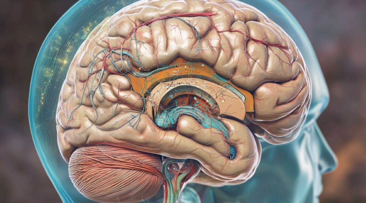
Summary: A recent study offers new insights into how brain regions coordinate during rest, using resting-state fMRI ( rsfMRI ) and neural recordings in mice. Researchers discovered that some mental activity is still “invisible” in conventional rsfMRI scans by comparing blood flow patterns with immediate neural activity. This obscure behavior suggests that crucial neurological behavior may not be captured by recent brain imaging techniques.
The findings, probably relevant to human studies, may enhance our knowledge of brain networks. Further study might improve the accuracy of interpreting mental exercise.
Important Information:
- rsfMRI measures mind exercise by tracking blood flow changes but does lose “invisible” neurological signals.
- Continuous neurological recordings revealed spatial and temporal alignment issues with rsfMRI.
- Results suggest a new element in mind activity interpretation, probably aiding animal studies.
Origin: Penn State
Researchers use a technique known as resting-state functional magnetic resonance imaging ( RSFMRI ) to understand how various brain regions interact with one another. RSFMRI does not explain how changes in blood circulation to various brain regions relate to what is happening with brain cells, which are organisms that send and receive messages in the form of digital signals. The process measures mental activity by monitoring changes in blood circulation to various brain regions.  ,  ,
The Dorothy Foehr Huck and J. Lloyd Huck Chair in Brain Imaging and professor of biological engineering at Penn State, Nanyin Zhang, a team of researchers led by and professor of biomedical engineering at Penn State, set out to answer this query. They just published their findings, made in mice, in the journal , covering.  ,  ,
To find out more about Zhang’s findings, Penn State News spoke with him. He is also connected to the departments of electronic engineering, engineering technology, and mechanics, as well as the Huck Institutes of the Life Sciences.  ,
Q: How does rsfMRI job? What can it show experts, and what are its limits?  ,
Zhang:  , Resting-state functional magnetic resonance imaging ( rsfMRI ) allows scientists to study how different parts of the brain work together. By examining their impromptu changes in blood circulation, this technique reveals when various brain regions are effective together.
We call these coordinated patterns “resting-state brain networks” ( RSNs ). We still do n’t fully understand how these blood flow changes are related to what is happening with the brain’s neural activities, despite the fact that we frequently use these RSNs. Lacking this understanding highlights a substantial difference in how efficient brain networks are understood.  ,
Q: How did you handle the limits of rsfMRI?  ,  ,
Zhang:  , We aimed to address the question of how RSNs and rsfMRI relate to unplanned neural activity. We employed a method that allows continuous monitoring of both rsfMRI and physiology signals, allowing the same brain site to measure both rsfMRI and rsfMRI signals at once.
We wanted to discover how neurological activities are directly related to unplanned blood flow changes in the brain by measuring these two signals simultaneously.  ,
Q: What were your results? What does this new knowledge about “invisible” signaling tell us?  ,
Zhang:  , We found a gap in the spatial and temporal relationships between the physiology message, which immediately measures neurological action, and the rsfMRI sign.
The electrophysiology signal can recapture the spatial patterns of brain-wide RSN connectivity that the rsfMRI signal reveals. However, these two types of signals do not align well over time.  ,  ,
These ostensibly contradictory findings raise the possibility that the rsfMRI signal is being influenced by electrophysiology’s “invisible signals,” which supports the current understanding of how neural activity and rsfMRI signal are related.
The conventional view believes the electrophysiology signal underlies most of the rsfMRI signal, whereas , our results suggest that a major source of the rsfMRI signal might actually originate from an electrophysiology-invisible component.  ,  ,  ,
Q: What implications do these findings have for studying brains and brain imaging?  ,  ,
Zhang: It is generally believed that the electrophysiology signal can be used to explain the rsfMRI signal. Our understanding of the neural basis of the rsfMRI signal, and thus the interpretation of RSNs, may not be sufficient due to the possibility that RSNs may result from electrophysiology-invisible brain activities that play a significant role in controlling rsfMRI signals.
To ensure that we are accurately interpreting brain activity, it is crucial to keep looking into the neural foundation of the rsfMRI signal.  ,  ,
Q: How does human research benefit from rsfMRI in animals?  ,
Zhang:  , The neural mechanism of rsfMRI in animals is very likely the same as that in humans. The current study’s findings may have significant implications for human rsfMRI studies.  ,
This work was supported by the National Institute of Mental Health and the National Institute of Neurological Disorders and Stroke.  ,
About this news story about brain mapping and neuroimaging
Author: Sarah Small
Source: Penn State
Contact: Sarah Small – Penn State
Image: The image is credited to Neuroscience News
Original Research: Open access.
Nanyin Zhang and colleagues ‘” Temporal and spatial differences between resting-state electrophysiological and fMRI signals.” eLife
Abstract
Temporal and spatial differences between resting-state electrophysiological and fMRI signals
Resting-state brain networks ( RSNs ) have been widely used in both medical and surgical settings, but it is unclear how to interpret them in terms of neural activity.
In two distinct brain regions of rats, we simultaneously recorded whole-brain resting-state functional magnetic resonance imaging ( rsfMRI ) and electrophysiology signals to address this crucial question.
Our research shows that spatial maps created by band-specific local field potential ( LFP ) power can account for up to 90 % of the spatial variation in RSNs created by rsfMRI signals for both recording sites.
Surprisingly, the local rsfMRI time course from the same location can only account for up to 35 % of the local rsfMRI time course’s temporal variance. The spatial patterns of rsfMRI-based RSNs are largely unaffected by regressing out the time series of LFP power from rsfMRI signals.
This disparity between resting-state electrophysiology and rsfMRI signals suggests that an underlying mechanism for the existence of an rsfMRI component is the absence of fully explained electrophysiological activity alone, leading to the existence of an ‘electrophysiology-invisible ‘ signal.
These findings provide a novel understanding of how RSN interpretation is interpreted.
