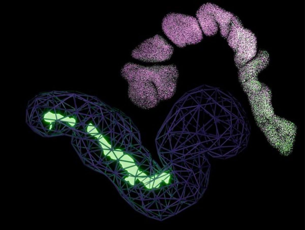
Summary: Academics have created human stem cell models that include the notochord, a crucial organ that controls the development of the bone and nervous system during embryogenesis.
The researchers were able to coerce human stem cells to form a notochord and a small trunk-like structure by using specific chemical signals inspired by biological processes in poultry, mouse, and monkey embryos. This creation, which reflects important aspects of human development, may help to advance research into spine-related birth defects and conditions like lumbar disk degeneration.
Important Facts:
- Notochord Role: The notochord acts as a” GPS” in embryos, guiding spine and nervous system development.
- Lab-Grown Trunk: Professionals recreated notochord muscle and a trunk-like architecture with organized neurological and bone tissues.
- Clinical Potential: The unit may help in studying baby defects and back problems linked to lumbar disc degradation.
Origin: Francis Crick Institute
For the first time, a tissue in the developing embryo acts like a navigation system, directing cells to construct the spine and nervous system ( the trunk ), has been created by researchers at the Francis Crick Institute.
The job, published today in , Nature, marks a major step forward in our ability to study how the human body takes design during early growth.
The notochord, a rod-shaped cells, is a vital part of the gallows of the developing brain. It is a distinguishing quality of all backboned animals and plays a crucial part in organizing the cells in the developing embryo.
Despite its importance, the difficulty of the architecture has meant it has been missing in past lab-grown models of human tree growth.
The scientists conducted the first analysis of chicken embryos to understand how the notochord normally forms. They were able to determine the schedule and series of the chemical signals required to create notochord tissue by comparing this to previously published information from mouse and monkey embryos.
With this framework, they created a specific sequence of chemical signs and used them to pull people stem cells into forming a notochord.
The hinge cells formed a small’ trunk-like’ framework, which naturally elongated to 1-2 millimetres in size. It contained tooth stem cells and developing neural cells in a manner that resembles that of human embryo development. This suggested that the notochord was encouraging tissues to develop the appropriate muscle at the appropriate time.
The research, according to the researchers, may provide insight into the study of spinal cord and back defects. It might also offer insight into the issues that affect the shock-absorbing seats between vertebrae created by the notochord, or intervertebral discs. When these cylinders degenerate with period, they can cause back pain.
James Briscoe, Group Leader of the Developmental Dynamics Laboratory, and senior author of the study, said:” The notochord acts like a GPS for the developing blastocyst, helping to establish the body’s primary shaft and guiding the formation of the bone and nervous system.
It’s been challenging to produce this essential tissues in the facility, which has so far limited our ability to study human development and problems. This opens windows to studying development circumstances that we’ve been in the dark about then that we’ve created a model that works.
Finding the precise chemistry signals that generate notochord was like finding the right meal, according to Tiago Rito, Postdoctoral Fellow in the Developmental Dynamics Laboratory and first author of the study. Past attempts to grow the notochord in the test may have failed because we had no idea how much time was needed to add the ingredients.
” The notochord in our lab-grown buildings appears to function similarly to how it would in a developing embryo,” says the researcher. It generates chemical signals that aid in the organization of surrounding tissues, just as it would during normal development.
About this information from neurodevelopment and genetics
Author: Clare Green
Source: Francis Crick Institute
Contact: Clare Green – Francis Crick Institute
Image: The image is credited to Tiago Rito
Original Research: Start exposure.
” Fast TGFβ signal restriction induces notochord” by Tiago Rito et cetera. Character
Abstract
Fast TGFβ signalling suppression induces notochord
The production of trunk tissues from forebears located in the lateral of the embryo is a key factor in the formation of the animal body.
Although in vivo models using embryonic stem cell replicate elements of this procedure, they lack essential components, most notably the notochord, a defining characteristic of chordates that resembles tissues.
Accordingly, cell types reliant on notochord signals are absent from recent models of human tree development.
To map the spatial organization of molecularly specific progenitor populations, we performed single-cell transcriptional analysis of chic embryos.
We investigated how separating individual pluripotent stem cells organized their trunk cell types according to the diagram.
We discovered that WNT pathway activation and induced expression of TBXT ( also known as BRA ) in combination with FGF-mediated MAPK signaling. Additionally, the proportions of various tissues sorts, including notochordal cells, were influenced by the proper suppression of WNT-induced NODAL and BMP signaling.
This made it possible to create a three-dimensional model of human stem development that exhibits morphogenetic motions, producing extended structures with lateral neural and mesodermal tissues and a notochord.
Our findings help to clarify the mechanisms that underlie the notochord formation in animal tissues and create a more complete in-vitro model of human trunk development. This opens the door for more detailed investigations of cells structuring in a context with physiological relevance.


