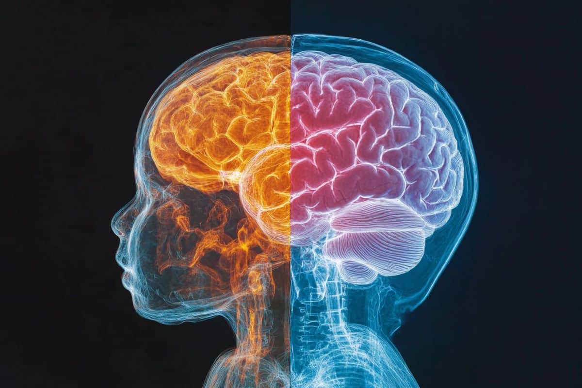
Summary: A research that looked at over 500 newborns found that there are mind construction differences between male and female infants from beginning. Females had more dark matter in areas relating to storage and feeling, whereas males had more white issue and dark matter in sensory and motor regions, whereas male brains were generally larger.
These findings point to the interaction between economic factors and gender differences in the mind during prenatal development. Experts emphasize that these differences are statistics and do not apply to everyone, underscoring the value of understanding diversity rather than giving significance to these differences.
Important Facts:
- First Sex Variations: Male and female head disparities are evident at birth, suggesting prenatal causes.
- Distinct Brain Structures: Women show more dark matter in memory and mental places, while males show more bright issue and sensory-motor dark matter.
- Neurodiversity Insight: Findings contribute to understanding problems like autism, diagnosed more often in men.
Origin: University of Cambridge
According to research from the University of Cambridge’s Autism Research Centre, gender differences in brain structure are existing from beginning.
When adjusted for full mind volume, sexual infants typically had significantly more gray matter than female brains, whereas male brains typically had significantly more light matter.
Grey matter is made up of nerve cell bodies and dendrites and is responsible for processing and interpreting data, such as pain, view, learning, talk, and cognition. White matter is composed of neurons, which are lengthy muscle fibers that connect cells from various parts of the brain.  ,
Yumnah Khan, a PhD student at the Autism Research Centre, who led the study, said:” Our investigation settles an age-old issue of whether male and female brains differ at birth. Although we are aware of differences between older children and older adults ‘ brains, our studies indicate that they are already existing in the earliest stages of life.
Because these gender differences are present right away after delivery, they may in part reflect biological gender differences during antenatal brain growth, which interact with environmental experiences over time to influence more sex differences in the brain.
The size of the sample has historically been a problem for research in this area. The Cambridge team attempted to address this by analyzing data from the Developing Human Connectome Project, which provides an MRI brain scan for infants soon after birth.
Having over 500 newborn babies in the study means that, statistically, the sample is ideal for detecting sex differences if they are present.
A second issue is whether any sex differences observed could be the result of other factors, such as body size differences.  ,
The Cambridge team discovered that male infants typically had significantly larger brains than females, even after gender differences in birth weight were taken into account.
After taking into account this disparity in total brain volume, females typically displayed larger volumes in grey matter relating to memory and emotional regulation, while males typically displayed larger volumes in grey matter relating to sensory processing and motor control at a regional level.
The findings of the study, the largest to date to investigate this question, are published in the journal , Biology of Sex Differences.
This is the largest study to date, according to Dr. Alex Tsompanidis, who oversaw the study.” We made sure that these differences are specific to the brain and not caused by general sex differences,” he said.
” To understand why males and females show differences in their relative grey and white matter volume, we are now studying the conditions of the prenatal environment, using population birth records, as well as , in vitro , cellular models of the developing brain.
This will enable us to compare the progression of male and female pregnancy and to understand whether specific biological factors, such as hormones or the placenta, are contributing to the differences we see in the brain.
The researchers point out that the differences between men and women are just average differences.
Dr Carrie Allison, Deputy Director of the Autism Research Centre, said:” The differences we see do not apply to all males or all females, but are only seen when you compare groups of males and females together. There is a lot a variation within, and a lot of overlap between, each group. ”  ,
Professor Simon Baron-Cohen, Director of the Autism Research Centre, added:” These differences do not imply the brains of males and females are better or worse. It’s just one example of neurodiversity.
Because males are more frequently diagnosed with autism, this research may be useful in understanding other types of neurodiversity, such as the brain in children who are later identified as such.
Funding: The research was funded by Cambridge University Development and Research, Trinity College, Cambridge, the Cambridge Trust, and the Simons Foundation Autism Research Initiative.
About this information on brain mapping and neurodevelopment
Author: Craig Brierley
Source: University of Cambridge
Contact: Craig Brierley – University of Cambridge
Image: The image is credited to Neuroscience News
Original Research: Open access.
Yumnah Khan and others ‘” At birth, there are sex differences in the human brain structure..” Biology of Sex Differences
Abstract
At birth, there are sex differences in the human brain structure.
Background
Sex differences in human brain anatomy have been well-documented, though remain significantly underexplored during early development. The neonatal period offers important insights into the influence of prenatal and early postnatal factors on sex differences in the brain.
Methods
Here, we assessed on-average sex differences in global and regional brain volumes in 514 newborns aged 0–28 , days ( 236 birth-assigned females and 278 birth-assigned males ) using data from the developing Human Connectome Project. To investigate sex differences in early postnatal brain development, we also looked into sex-by-age interactions.
Results
On average, males had significantly larger intracranial and total brain volumes, even after controlling for birth weight. Females had significantly higher total cortical gray matter volumes after controlling for total brain volume, whereas males had higher total white matter volumes.
Females had significantly higher white matter volumes in the corpus callosum and higher gray matter volumes in the bilateral parahippocampal gyri ( posterior parts ), left anterior cingulate gyri, bilateral parietal lobes, and left caudate nucleus after controlling for total brain volume in regional comparisons.
Males ‘ right medial, inferior temporal, post-posterior, and right subthalamic gyruses both had significantly higher gray matter volumes.
Effect sizes ranged from small for regional comparisons to large for global comparisons. The left superior temporal gyrus and the left anterior cingulate ( posterior parts ) both experienced significant sex-by-age interactions.
Conclusions
Our findings demonstrate that prenatal factors play a significant role in shaping sex differences in the brain, demonstrating that sex differences in brain structure are already present at birth and remain aproximally stable during early postnatal development.


