Summary: A fresh AI unit can determine how quickly a person’s head is aging using MRI images, providing a strong tool for detecting cognitive drop. This concept tracks brain aging over time, identifying the areas that are most damaged and relating changes to mental function, in contrast to previous methods.
Early treatment might help prevent degenerative diseases, according to research that found that brain aging has a strong correlation with cognitive impairment. This discovery could lead to better diagnostics, personal remedies, and earlier verification of Alzheimer’s risk.
Major Information
- The AI type uses MRI scans over time to determine how quickly the head years, providing a more precise method than earlier methods.
- Mental Decline Predictor: Better mind aging correlates with decreased mental function, including slower running speed and memory decline.
- Potential for Early Diagnosis: The unit may help identify people at high risk for Alzheimer’s before symptoms appear, enabling earlier initiatives.
Origin: USC
A new artificial intelligence type measures how quickly a patient’s mind is ageing and could be a powerful new instrument for knowing, preventing and treating mental decline and memory, according to USC researchers.
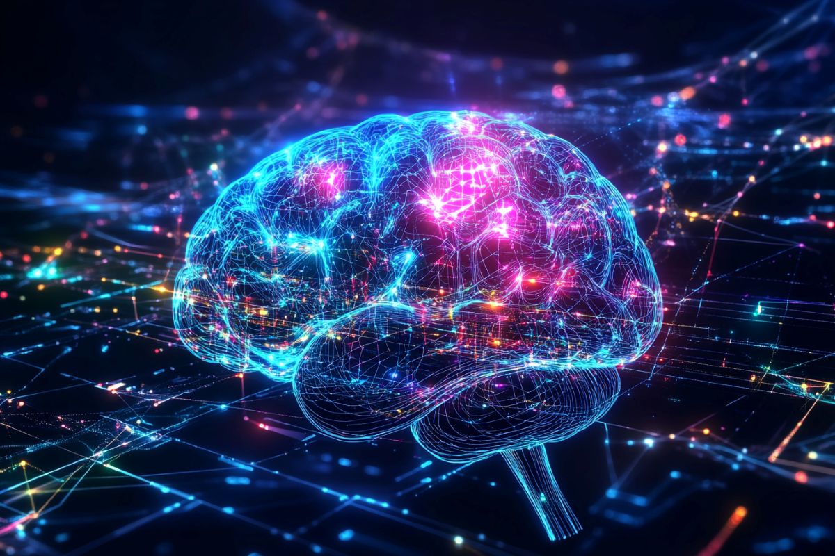
The first-of-its-kind tool can non-invasively track the pace of brain changes by analyzing magnetic resonance imaging ( MRI ) scans.
Better brain aging closely correlates with a higher risk of cognitive deficits, said , Andrei Irimia, associate teacher of gerontology, biomedical engineering, statistical &, mathematical biology and neuroscience at the USC Leonard Davis School of Gerontology and visiting associate professor of internal medicine at King’s College London.
This is a novel test that could alter how we monitor brain health in the clinic as well as in the research lab,” he said. ” Knowing how fast one’s brain is aging can be powerful”.
Irimia is the senior author of the study, which details the new model’s predictive power and its description. It was published on February 24, 2025 in the Proceedings of the National Academy of Sciences.
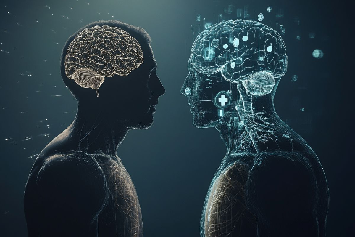
chronological versus biological brain age
Biological age is distinct from an individual’s chronological age, Irimia said. Due to how well their bodies are working and how “old” the body’s tissues appear at a cellular level, two people who are the same age can have very different biological ages.
Blood samples are used frequently to measure epigenetic aging and DNA methylation, which affect the roles of genes in the cell. However, measuring biological age from blood samples is a poor strategy for measuring the brain’s age, Irimia explained.
Blood cells entering the brain cannot cross the bloodstream, preventing a blood sample from one’s arm from accurately displaying methylation and other aging-related processes in the brain.
In contrast, it is much more expensive to obtain a sample directly from a patient’s brain, making it impossible to measure DNA methylation and other brain aging parameters directly from living human brain cells.
The potential of MRI scans to non-invasively assess the biological age of the brain was highlighted by recent research by Irimia  and colleagues.
To compare a patient’s brain anatomy to data gathered from thousands of people of various ages and cognitive health outcomes, the earlier model used AI analysis.
However, the cross-sectional nature of analyzing one MRI scan to estimate brain age had major limitations, he said.
The previous model, for instance, couldn’t tell whether a patient’s brain was ten years “older” than their calendar age, nor could it tell whether that additional aging had taken place sooner or later in their lives.
A more accurate depiction of brain aging
A recently developed three-dimensional convolutional neural network ( 3D-CNN) provides a more precise method of determining brain age over time. The model was developed in collaboration with Paul Bogdan, associate professor of electrical and computer engineering and USC Viterbi School of Engineering’s Jack Munushian Early Career Chair.
Unlike traditional cross-sectional approaches, which estimate brain age from one scan at a single time point, this longitudinal method compares baseline and follow-up MRI scans from the same individual. It therefore more precisely detects neuroanatomical changes related to accelerated or decelerated aging.
According to Bogdan, the 3D-CNN also produces interpretable” saliency maps,” which show which brain regions are most crucial for determining the rate of aging.
The new model’s calculations of brain aging speed were sensitively correlated with changes in cognitive function tests administered to a group of 104 cognitively healthy adults and 140 patients with Alzheimer’s disease when applied to a group of 140 cognitively healthy adults.
According to Bogdan,” The alignment of these measures with cognitive test results indicates that the framework may serve as an early biomarker of neurocognitive decline.” Additionally, it demonstrates its applicability in cognitively normal people as well as those who have cognitive impairment.
He added that the model could be used to determine which treatments would be most effective based on individual characteristics and have the potential to better characterize both healthy aging and disease trajectories.
According to Irimia,” the rates of brain aging are significantly related to changes in cognitive function.” ” So, if you have a high rate of brain aging, you’re more likely to have a high rate of degradation in cognitive function, including memory, executive speed, executive function, and processing speed. The changes we see in the anatomy are related to the changes we see in these people’s cognition, not just an anatomic measure.
Looking ahead
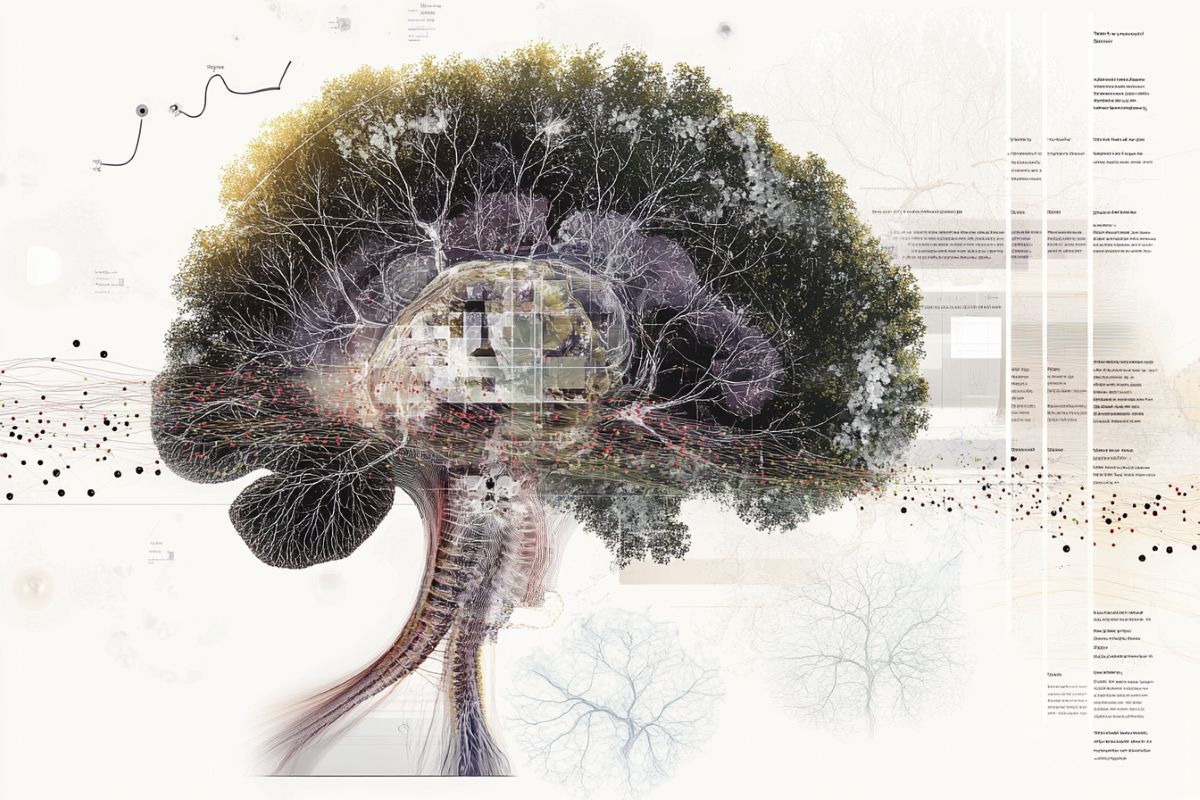
Irimia and coauthors also point out in the study how the new model was able to distinguish different ages at various brain regions. Understanding these variations, including how they vary depending on genetics, environment, and lifestyle factors, might shed light on how various pathologies develop in the brain, according to Irimia.
The study also revealed that different regions of the brain ages at different rates, which might explain why men and women are at risk for neurodegenerative diseases, including Alzheimer’s, he added.
Irimia added that he is also excited about the potential for the new model to identify those who have more advanced cognitive impairment before they exhibit any symptoms of cognitive impairment.
Although new medications for treating Alzheimer’s disease have been developed, their efficacy has been lower than researchers and doctors had anticipated, possibly because patients may not be starting the medication until there is already a significant amount of Alzheimer’s pathology in the brain, he explained.
” One thing that my lab is very interested in is estimating risk for Alzheimer’s, we’d like to one day be able to say,’ Right now, it looks like this person has a 30 % risk for Alzheimer’s.’ We’re not there yet, but we’re working on it”, Irimia said.
” I believe this kind of measurement will be very useful in creating prognostic variables that can be used to make Alzheimer’s risk predictions. That would be really powerful, especially as we start developing potential drugs for prevention”.
Along with Irimia and Bogdan, the study’s authors included first author Chenzhong Yin and Heng Ping of the USC Viterbi School of Engineering and Phoebe E. Imms, Nahian F. Chowdhury, Nikhil N. Chaudhari, and Haoqing Wang of the USC Leonard Davis School of Gerontology.
Funding: Support for the study came from the National Institutes of Health ( NIH) under grants R01 NS 100973, RF1 AG 082201, and R01 AG 079957, the Department of Defense under contract W81XWH-18-1-0413, the National Science Foundation under CAREER Award CPS/CNS-1453860, grants MCB-1936775 and CNS-1932620, the U. S. Army Research Office under grant W911NF-23-1-0111, DARPA under a Young Faculty Award and under Director Award N66001-17-1-4044, an Intel Faculty Award, Northtrop Grumman, the Hanson-Thorell Research Scholarship Fund, the Undergraduate Research Associate Program, the Center for Undergraduate Research in Viterbi Engineering ( CURVE ) at USC, and anonymous donors.
About this news about research into AI and brain aging
Author: Elizabeth Newcomb
Source: USC
Contact: Elizabeth Newcomb – USC
Image: The image is credited to Neuroscience News
Original Research: Closed access.
By Andrei Irimia and colleagues,” Deep learning to quantify the rate of brain aging in relation to neurocognitive changes.” PNAS
Abstract
Deep learning to analyze how quickly the brain ages in relation to neurocognitive changes
MRIs can be used to estimate brain age ( BA ), a different from chronological age ( CA ), to compare neuroanatomic aging in cognitively normal ( CN) individuals. BA, however, is a cross-sectional measure that summarizes cumulative neuroanatomic aging since birth.
Thus, it conveys poorly recent or contemporaneous aging trends, which can be better quantified by the (temporal ) pace , P , of brain aging. Many approaches to map , P, however, rely on quantifying DNA methylation in whole-blood cells, which the blood–brain barrier separates from neural brain cells.
We introduce a three-dimensional convolutional neural network ( 3D-CNN) to estimate , P , noninvasively from longitudinal MRI.
Our longitudinal model ( LM) is trained on MRIs from 2, 055 CN adults, validated in 1, 304 CN adults, and further applied to an independent cohort of 104 CN adults and 140 patients with Alzheimer’s disease ( AD ). In its test set, the LM computes , P , with a mean absolute error ( MAE ) of 0.16 y ( 7 % mean error ).
This significantly outperforms the most accurate cross-sectional model, whose MAE of 1.85 y has 83 % error. We map anatomic variations in regional brain aging rates that vary depending on sex, decade of life, and neurocognitive status by combining the LM with an interpretable CNN saliency approach.
LM estimates of , P , are significantly associated with changes in cognitive functioning across domains. This underscores the LM’s ability to estimate , P , in a way that captures the relationship between neuroanatomic and neurocognitive aging.
This study builds on earlier methods for assessing AD risk that estimate how many people’s rates of adverse cognitive change as they age.