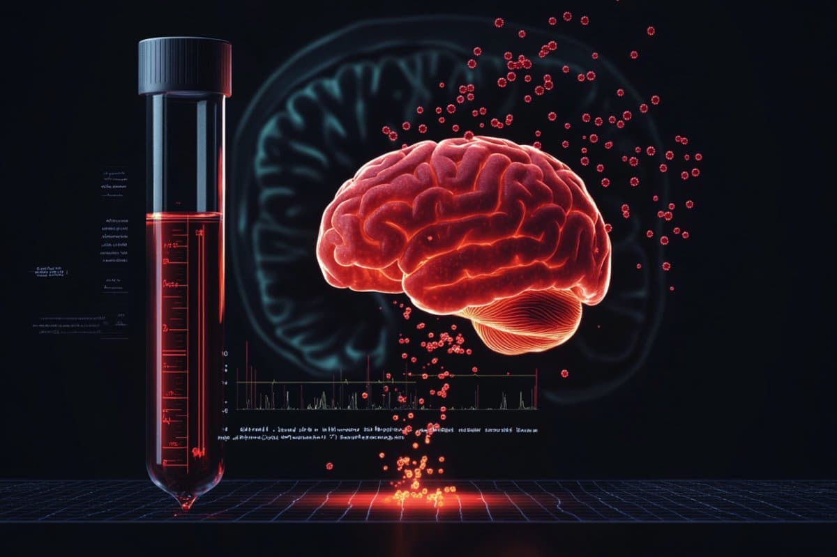
Summary: Researchers have created a personalized blood test that may provide a quicker, less intrusive way to monitor the progression of high-grade brain. The test may identify tumor DNA in the brain actually before modifications appear on MRI scans by identifying distinct DNA crossings from each victim’s tumor.
These DNA fragments provide a delicate early alert for the spread of disease by breaking some of the blood-brain challenge. In 93 % of cases, the test revealed tumor activity, which suggests promise as a diagnostic tool for adjusting treatments and tracking results in real time.
Important Information:
- Focus on DNA Junctions: Experts focused on tumor-specific DNA crossings, which are more prevalent and simple to identify than other fragments.
- First Detection: Blood tumor DNA levels occasionally increased before any MRI changes, giving earlier insights.
- High Precision: 93 % of people with known DNA junctions had tumor DNA confirmed by the personal evaluation.
Mayo Clinic as cause
Researchers at the Mayo Clinic have discovered a possible new method to track the growth of high-grade gliomas, one of the most extreme forms of brain cancer. Their research suggests that a personalized blood test that is individualized to each hospital’s tumor DNA could be used to determine whether the cancer is progressing more quickly and less in invasively.
To screen gliomas, clinicians now rely on scans and clinical biopsies, but both techniques have drawbacks. For instance, scans frequently fail to differentiate between tumor growth and treatment-related inflammation. Intrusive procedures are necessary for daily monitoring of biopsies, making them impractical.
This innovative method, which was published in the journal Clinical Cancer Research, does give professionals a new tool to monitor lesion changes over time and adjust treatment as needed.
The investigation focuses on blood-circulating lesion DNA bits. Some brain cells pass away as they grow, releasing genetic markers that are specific to the tumor and releasing fragments of their Genome into the bloodstream.
Compared to many other tumours, cancer release fewer DNA bits into the body. This is because of the blood-brain challenge, a healthy defense against some materials from leaving the mind.
Researchers concentrated on DNA junctions, a higher-presented sort of tumor-specific DNA piece, to conquer this impediment. Researchers were able to detect even the smallest indicators of tumor growth by targeting these markers, increasing their awareness.
These DNA crossings form when the tumor’s biological materials disintegrates and organizes, in contrast to regular DNA, which follows a structured format. These enhanced DNA junctions may provide a more accurate picture of the progression of disease, according to the study.
This research builds on years of research into genomic rearrangements and provides a more in-depth understanding of the chemical mechanisms causing gliomas, says lead author George Vasmatzis, Ph.D. Co-director of the Mayo Clinic Comprehensive Cancer Center, Mayo Clinic’s Center for Individualized Medicine, and Mayo Clinic’s Biomarker Discovery Program, Dr.
It opens up new avenues for targeted interventions and patient-specific tracking.
Scientists analyzed samples from patients with high-grade cancer for the purpose of the investigation. They identified patient-specific DNA intersections and used complete genome sequencing to map each tumor’s distinct biological template.
Finally, to look for these biological markers in blood, researchers created personalized body tests.
In the majority of the cases involving these DNA junctions, the test found lesion DNA. Before MRI scans revealed any modifications, tumor DNA amounts in some people rose, potentially giving rise to early warning signs of disease progression.
Dr. Vasmatzis and Terry Burns, M. D., Ph. D., connect cutting-edge research and clinical training. D., a neurosurgeon at Rochester, Minnesota’s Mayo Clinic, collaborated on the study.
By observing each tumor’s distinctive molecular signature, review co-author Dr. Burns says,” We’re aiming to switch from a reactive approach to one that’s much more proactive.”
” This study could provide the foundation for instruments that enable professionals to make the best decisions about treatments as soon as possible,” said Dr. B. S.
Future studies may examine whether the effectiveness of blood-based lesion tracking predicts the progression of glioma across a larger patient population.
About this information from studies into brain tumor and genetics
Author: Sharon Theimer
Source: Mayo Clinic
Contact: Sharon Theimer – Mayo Clinic
Image: The image is credited to Neuroscience News
Original Research: Disclosed entry.
George Vasmatzis and colleagues ‘” Personal Tumor-Specific Enhanced DNA Junctions in Peripheral Blood of Patients with High-Grade Gliomas” Cancer Research in Clinical Settings
Abstract
High-grade gliomas patients have individualized personalized personal enhanced DNA junctions in their peripheral blood.
Purpose:
Due to treatment-related changes in imaging and the need for neurosurgical intervention to obtain diagnostic tissue, it is challenging to monitor disease progression in patients with high-grade gliomas ( HGG ). In HGG, oncogenes are frequently amplified by DNA junctions, which could increase the number of these DNA fragments in body compared to monoallelic mutations.
We used patient-specific DNA intersections associated with oncogene capacitances to test a cell-free DNA method for disease diagnosis in the blood of patients with HGG in this study.
Design for experimentation:
Grade 3 or 4 isocitrate dehydrogenase–mutant or wild-type astrocytomas were identified using whole-genome decoding. Using patient-specific primer specifically designed for the enhanced bridge, individualized qPCR assays were created. These intersections were found in individual blood samples at ctDNA levels.
Results:
In 18 patients with tumor-associated primary amplification, 15 had tumor-associated primary amplification, and three had tumor-associated focus amplifications, individualized semi-qPCR assays were used to evaluate unique amplified junctions in presurgical plasma. No controls or 14 of 15 (93.3 % ) patients with amplified junctions had high copy-number junctions in their plasma.
In sequential blood samples from five patients, changes in junction abundance were correlated with illness trajectory, with the most abundant enhanced junctions occurring prior to radiographic disease progression.
Conclusions:
Patient-specific enhanced junctions were safely found in the majority of the patients who had quality 3 or 4 astrocytomas and had tumor-associated amplifications. Plasma examples ‘ vertical analysis revealed a link between cytoreduction and development and the disease’s progression.


