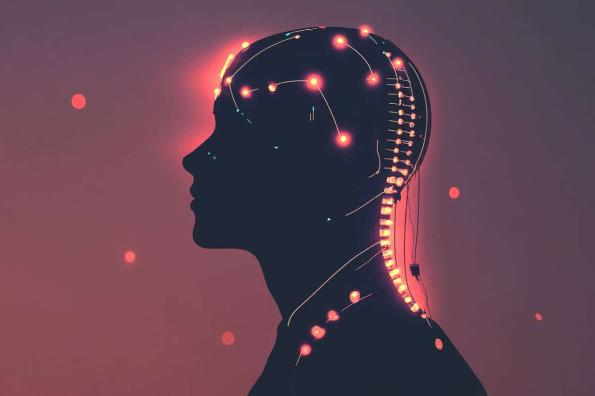
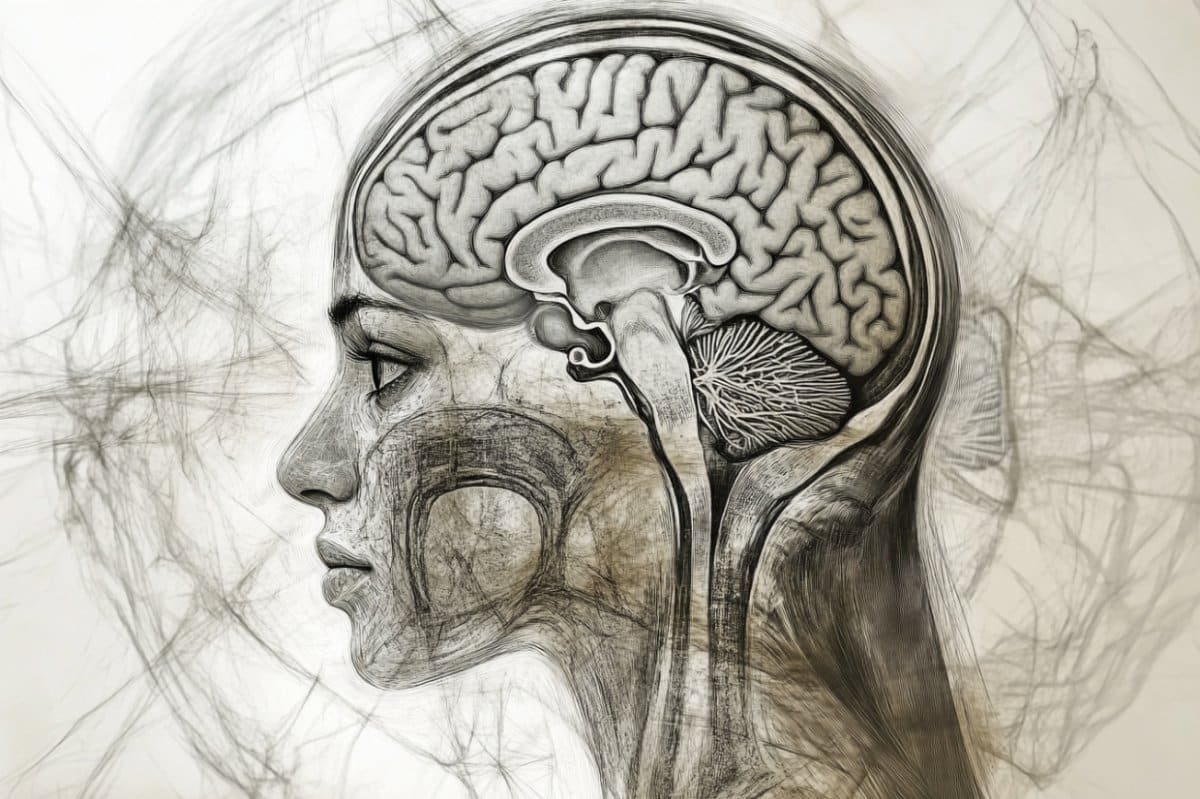
Summary: Researchers have developed a invasive brain-spine software that detects motion intentions and cues spinal cord stimulus to aid treatment. Using EEG cap, they trained a receiver to understand when individuals either moved or imagined moving their feet.
This proof-of-concept research shows that imagined activity alone can effectively cause lumbar excitement, opening new possibilities for restoring deliberate movement after spinal cord injury. Coming efforts aim to create widespread decoders to simplify medical application.
Important Information:
- Exercise Intention Decoded: A invasive EEG receiver accurately predicts calf movement or imagined movement.
- Lumbar Stimulation Triggered: Mind signals can signal spinal cord stimulation to strengthen volunteer movement.
- Clinical Potential: This approach may lead to percutaneous treatment therapies for people with spinal cord injuries.
Origin: WUSTL
When a man sustains an injury to the spinal cord, the regular contact between the mind and the lumbar wires below the injuries are interrupted, resulting in immobility.
Because the head is functioning normally, as is the spinal cord below the wound, researchers have been working to re-establish the conversation to allow for treatment and possible recover movement.
Ismael Seáñez, assistant professor of biomedical engineering in the McKelvey School of Engineering at Washington University in St. Louis and of neurosurgery at WashU Medicine, and members of his lab, including Carolyn Atkinson, a doctoral student, have developed a type of decoder to restore that communication.
Through experiments in their lab with 17 human subjects without a spinal cord injury, they were able to cue movement in the lower leg with transcutaneous spinal cord stimulation, or noninvasive, external electrical pulses.
Results of the research were published online April 25, 2025, in the Journal of NeuroEngineering and Rehabilitation.
The team used a special cap fitted with noninvasive electrodes that measure brain activity through electroencephalography ( EEG).
While wearing the cap, seated volunteers were asked to extend their leg at the knee, then to only think about extending their leg– while keeping it still — so researchers could record the brain waves in both exercises.
The team provided the neural activity to the decoder, or algorithm, so it could learn how the brain waves act in both circumstances. They found that the actual movement and imagined movement used similar neural strategies.
“After we give the decoder this data, it learns to predict based on neural activity whenever there is movement or no movement, ” Seáñez said. “We show that we can predict whenever someone is thinking about moving their leg, even if their leg does not actually move. ”
The team used controls to ensure that the volunteers were truly imagining movement and not actually moving.
“ Whenever people move, this can introduce signal noise, and we want to make sure that the signal noise is not what we’re learning to predict, ” Seáñez said.
“It’s movement intention or brain activity that we want to predict, so we have people imagine that they’re extending their leg and use the same algorithm that has been trained on people moving to predict whether they were imagining or not. ”
Seáñez said this reveals two things.
“One, that it ’s more likely that we’re decoding movement intention and not an artifact, or noise, and second, whenever we employ this on people with spinal cord injury who will not have that ability to actually move their legs for us to label the data, we could use their imagination of moving a leg to train our decoder. ”
Seáñez said the proof-of-concept study is a first step toward developing a noninvasive brain-spine interface that uses real-time predictions to deliver transcutaneous spinal cord stimulation to reinforce voluntary movement in a single joint in rehabilitation in patients with a spinal cord injury.
Going forward, the team plans to test a generalized decoder trained on data from all participants that could determine whether a universal decoder could perform as well as a personalized one and simplify its use in clinical settings.
Funding: Funding for this research was provided by the McDonnell Center for Systems Neuroscience at Washington University in St. Louis; the National Institutes of Health ( K12-HD073945, K01-NS127936; R01-EB026439; P41-EB018783 ); the Department of Biomedical Engineering in McKelvey Engineering at WashU; and the Department of Neurosurgery at WashU Medicine.
About this SCI and neurotech research news
Author: Leah Shaffer
Source: WUSTL
Contact: Leah Shaffer – WUSTL
Image: The image is credited to Neuroscience News
Original Research: Open access.
“Development and evaluation of a non-invasive brain-spine interface using transcutaneous spinal cord stimulation ” by Carolyn Atkinson et al. Journal of NeuroEngineering and Rehabilitation
Abstract
Development and evaluation of a non-invasive brain-spine interface using transcutaneous spinal cord stimulation
Motor rehabilitation is a therapeutic process to facilitate functional recovery in people with spinal cord injury (SCI). However, its efficacy is limited to areas with remaining sensorimotor function.
Spinal cord stimulation (SCS) creates a temporary prosthetic effect that may allow further rehabilitation-induced recovery in individuals without remaining sensorimotor function, thereby extending the therapeutic reach of motor rehabilitation to individuals with more severe injuries.
In this work, we report our first steps in developing a non-invasive brain-spine interface ( BSI) based on electroencephalography ( EEG ) and transcutaneous spinal cord stimulation ( tSCS).
The objective of this study was to identify EEG-based neural correlates of lower limb movement in the sensorimotor cortex of unimpaired individuals ( N = 17 ) and to quantify the performance of a linear discriminant analysis ( LDA ) decoder in detecting movement onset from these neural correlates.
Our results show that initiation of knee extension was associated with event-related desynchronization in the central-medial cortical regions at frequency bands between 4 and 44 Hz.
Our neural decoder using µ ( 8–12 Hz ), low β ( 16–20 Hz ), and high β ( 24–28 Hz ) frequency bands achieved an average area under the curve ( AUC) of 0. 83 ± 0. 06 s. d. ( n = 7 ) during a cued movement task offline.
Generalization to imagery and uncued movement tasks served as positive controls to verify robustness against movement artifacts and cue-related confounds, respectively.
With the addition of real-time decoder-modulated tSCS, the neural decoder performed with an average AUC of 0. 81 ± 0. 05 s. d. ( n = 9 ) on cued movement and 0. 68 ± 0. 12 s. d. ( n = 9 ) on uncued movement.
Our results suggest that the decrease in decoder performance in uncued movement may be due to differences in underlying cortical strategies between conditions.
Furthermore, we explore alternative applications of the BSI system by testing neural decoders trained on uncued movement and imagery tasks.
By developing a non-invasive BSI, tSCS can be timed to be delivered only during voluntary effort, which may have implications for improving rehabilitation.


