Summary: New research has revealed that visual brain neurons can adapt to difficult stimuli more quickly than originally believed, resulting in dynamic response to complex stimuli during thing recognition tasks. While physical running has traditionally been viewed as largely proposes, this research shows that feedback from higher brain areas conveys previous knowledge and behavioural context to form perception.
This” top-down” details enables quick sensory neurons to gradually alter their response based on the task at hand. These findings challenge conventional theories of physical control and may have an impact on how to understand understanding and brain disorders like adhd.
Important Facts:
- Quick sensory neurons can quickly adapt to new tasks and previous experiences.
- Top-Down Feedback: Lower aesthetic areas receive cultural information to aid in perception.
- Relevance for Autism: Understanding feedback methods may shed light on visual variations in autism.
Rockefeller University is the cause.
From the moment we open our eyes, our brains begin making inside images of the world around us. We psychologically assemble pieces of scenes into recognized objects thanks to neurons in the physical brain.
The lateral aesthetic cerebral pathway, which extends from the front of the brain’s main visual cortex to the temporal lobes, is where this process takes place.
Long-standing theories have been made that certain neurons along this route handle particular kinds of information, depending on where they are located, and that feedforward, the way to the top of the visual cortical hierarchy, provides the most visible information.
Although it has long been known to occur in the opposite direction of cerebral connections, it is frequently referred to as feedback, its true purpose has not been well understood.
Ongoing analysis from the laboratory of Rockefeller University’s Charles D. Gilbert is revealing an essential role for input along the physical road.
This sway torrent carries so-called” best down” data across cerebral areas that is informed by our previous encounters with an item, as his team demonstrates in a recent paper , published , in , PNAS.
This flow has the effect that neurons in this route may change their responses at any given time to the info they are giving them.
” Even at the first phases of object perception, the cells are sensitive to much more complex visual impulses than had formerly been believed, and that potential is informed by input from higher cortical areas”, says Gilbert, mind of the , Laboratory of Neurobiology.
a different process
Gilbert’s lab has spent many years studying the brain’s fundamental mechanisms for understanding how information is organized.
” The classical view of this pathway proposes that neurons at its beginning can only perceive simple information such as a line segment, and that complexity increases the farther up the hierarchy you go until you reach the neurons that will only respond to a specific level of complexity”, he says.
His lab’s previous investigations point to the possibility that this view may be incorrect. For instance, his team has discovered that the visual cortex has the ability to alter its circuitry and functional characteristics, a trait known as plasticity.
And in work done with his Rockefeller colleague ( and Nobel Prize winner )  , Torsten N. Wiesel, Gilbert discovered long-range horizontal connections along cortical circuits, which enable neurons to link bits of information over much larger areas of the visual field than had been thought.
He’s also demonstrated that neurons can switch between inputs that are task-relevant and those that are task-unrelated, which highlights the dexterity of their functional characteristics.
We were attempting to establish that these capabilities are a part of our typical process of object recognition for the current study, he says.
Seeing is understanding
The animals ‘ lab, which Gilbert’s lab had spent several years working with, images of a variety of objects including fruits, vegetables, tools, and machines, for the purpose of studying a pair of macaques.
The researchers used fMRI to track the animals ‘ brain activity as they learned to recognize these objects and to identify which regions of their brains responded to the visual stimuli. ( This method was pioneered by Gilbert’s Rockefeller colleague Winrich Freiwald, who has used it to identify regions of the brain that are  , responsive to faces. )
The animals were shown images of the objects they had been trained to recognize, so they then implanted electrode arrays, which made it possible to track the activity of individual nerve cells.
Sometimes they were shown the entire object, and other times a partial or expertly cropped image. Then they were shown a variety of different visual stimuli and indicated whether they found a match to the original object or not.
Because there is a delay between when an object cue is seen and when a second object or object component is shown, to which they are trained to determine whether the second image corresponds to the initial cue, Gilbert calls these tasks “delayed match-to-sample tasks.”
They have to use their working memory to remember the original image while they’re looking through all the visual stimuli to find a match.
Adaptive processing
A single neuron may be more responsive to one target as opposed to a number of visual targets, according to the researchers ‘ findings, and with another cue, they’ll be more responsive to a different target.
We discovered that these neurons are adaptive processors that can perform various functions that are appropriate for the current behavioral context, Gilbert says.
They also demonstrated that the neurons found at the beginning of the pathway, thought to be limited to responding to simple visual information, were not actually so constrained in their abilities.
He claims that these neurons are much more tolerant to complex visual stimuli than what was previously believed.
There doesn’t seem to be as much of a difference in the degree of complexity in the higher cortical regions as previously thought.
These findings bolster what Gilbert believes is a novel view of cortical processing: that adult neurons do not have fixed functional properties but are instead dynamically tuned, changing their specificities with varying sensory experience.
Recipient feedback connections might have a potential functional role in object recognition because their dynamic capabilities are influenced by the flow of information from higher cortical regions to these lower ones.
We discovered that these so-called” top-down” feedback connections transmit information from areas of the visual cortex that represent previously stored data about the nature and identity of objects, which is made available through experience and behavioral context, according to him.
” In a sense, the higher-order cortical areas send an instruction to the lower areas to perform a particular calculation, and the return signal—the feedforward signal—is the result of that calculation.
As we recognize an object and, in general, make sense of our surroundings, these interactions are likely to be continuing.
applications for autism research
The findings are part of an increasing recognition of the importance and prevalence of feedback information flow in the visual cortex—and perhaps far beyond.
I think top-down interactions are essential to all brain functions, including motor control, higher order cognitive functions, and other senses, so understanding the cellular and circuitry that underlie these interactions could help us better understand the causes of brain disorders, according to Gilbert.
His lab is beginning to investigate autism in both behavioral and imaging levels. Will Snyder, a research specialist in Gilbert’s lab, will study perceptual differences between autism-model mice and their wild-type littermates.
The lab will also use the highly developed neuroimaging techniques in the Elizabeth R. Miller Brain Observatory, an interdisciplinary research center located on Rockefeller’s campus, to observe large neuronal populations in the animals ‘ brains as they engage in natural behaviors.
Our goal is to see if we can identify any perceptual differences between these two groups and the functioning of cortical circuits that might explain these differences, Gilbert says.
About this visual neuroscience research news
Author: Katherine Fenz
Source: Rockefeller University
Contact: Katherine Fenz – Rockefeller University
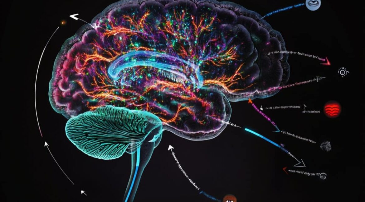
Image: The image is credited to Neuroscience News
Original Research: Private access.
” The ventral visual cortical pathway exhibits expectation-dependent stimulus selectivity.” by Charles D. Gilbert et al. PNAS
Abstract
The ventral visual cortical pathway exhibits expectation-dependent stimulus selectivity.
The hierarchical view of the ventral object recognition pathway is primarily based on feedforward mechanisms, starting from a fixed basis set of object primitives and ending on a representation of whole objects in the inferotemporal cortex.
Here, we offer a different viewpoint. Instead of being a fixed “labelled line” for a particular feature, neurons are constantly altering their stimulus selectivity based on a moment-to-moment basis, as dictated by top-down effects on object expectation and perceptual task.
Here, we also derive the selectivity for stimulus features from an ethologically curated stimulus set, based on a delayed match-to-sample task, that finds components that are informative for object recognition in addition to full objects, though the top–down effects were seen for both informative and uninformative components.
Functional MRI was used to identify cortical regions that respond to these stimuli to help place chronically implanted electrode arrays.
