Summary: Researchers have identified a particular subset of brain cells that act as appetite-reducers and weight-loss signals without producing side effects like discomfort. In mice, activating these cells caused a decrease in food consumption and fat loss, thereby reversing the product’s advantages.
In contrast, removing them reduced the weight-loss benefits of semaglutide, but not the negative results it produces. This finding might lead to more precise overweight treatments that preserve the advantages of GLP-1R agonists while reducing pain.
Important Information:
- Targeted Neurons: A particular group of cells in the lateral dopaminergic complex controls the appetite-suppressing ability of semaglutide.
- Side Impact Separation: These neurons are no significantly involved in side results like muscle loss or nausea.
- Medical Potential: Eliminating the need for these neurons may improve obesity treatments while reducing undesirable symptoms.
University of Gothenburg, cause
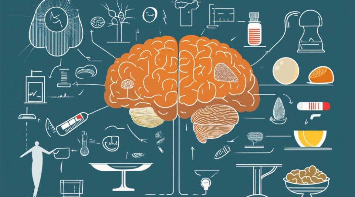
Without producing discomfort, a particular group of brain stem brain tissue appears to be in charge of how semaglutide affects hunger and weight.
The University of Gothenburg’s breakthrough may pave the way for improved treatments for obesity.
Semaglutide belongs to a class of medications known as GLP-1R receptors and has been shown to effectively lower body fat and food diet. Although the substance is already well established as a part of the cure for obesity and type 2 diabetes, it may have side effects like nausea and tissue damage.
Researchers at the University of Gothenburg’s Sahlgrenska Academy recently discovered that it is possible to distinguish between the brain’s brain cells that produce side effects, such as reduced food intake and large damage, from those that produce side effects in a new study.
The study has been published in the book Cell Metabolism.
Nerve tissue that have been activated
The scientists used mice to study how semaglutide affects the brain. They identified which nerve cells that the drug caused to activate, and they were able to promote these cells without using the medication directly.
The end result was that the animals lost weight and ate less, just as they did when semaglutide was administered. When these brain cells were removed, the drug’s impact on appetite and fat loss didn’t significantly decrease. But, discomfort and muscle loss persisted.
This suggests that these brain cells are in charge of the semaglutide’s useful results. We have thus identified a particular class of brain cells that are required for the weight and appetite-related effects of semaglutide, but which do not appear to be significantly responsible for side effects like nausea.
According to Jlia Teixidor-Deulofeu, primary author of the study and PhD student at Sahlgrenska Academy at the University of Gothenburg,” If we can target the treatment it, we may be able to maintain the positive effects while reducing side effects.”
The brain’s reaction
The lateral dopaminergic complex, which contains the identified nerve cells, is a region of the brain where it is located. The outcome is not only a first step toward possible improved treatment, but also a first step toward uncovering new information about how semaglutide functions in the brain.
Additionally, the research provides more information about how the brain stem controls our power balance.
Semaglutide and different GLP-1R agonists are already being prescribed to more and more people, and they are also being looked into for potential other uses, such as neurodegenerative diseases and substance use disorders.
” It is crucial to comprehend how these medicines actually function. The more we understand this, the more opportunities there are to improve them, according to Linda Engström Ruud, researcher and superintendent to PhD students Julian Blid Sköldheden and Jlia Teixidor-Deulofeu, both of whom worked on the project.
About this semaglutide and information from science study
Author: Jlia Teixidor-Deulofeu
Source: University of Gothenburg
Contact: Júlia Teixidor-Deulofeu – University of Gothenburg
Image: The image is credited to Neuroscience News
Open access to original analysis
Jlia Teixidor-Deulofeu et cetera.,” Semaglutide effects on power balance aremediated by neurons in the medial vagal complex.” Cell Digestion
Abstract
Cells in the medial dopaminergic complex, Adcyap1+, and semaglutide effects on the energy balance
The use of the GLP-1R receptor semaglutide has revolutionized the obesity treatment, but its molecular effects on energy balance are still obscure.
Here, we demonstrate that the drug’s effects of promoting fat usage and conditioned flavor aversion are mirrored by the reactivation of semaglutide-responsive lateral vagal difficult neurons.
We find that many of the semaglutide-activated area postrema ( AP ) and nucleus of the solitary tract (NTS ) neurons express , Adcyap1 , mRNA, and ablation of AP/NTS , Adcyap1+ , neurons largely reverse semaglutide’s effects on energy balance acutely in lean mice and subchronically treated
With just a small impact on controlled style dislike, semaglutide-activated AP/NTS , Adcyap1+ , cells promote the loss of fat more than muscular mass.
Additionally, NTS  and Adcyap1+  cells are required for the activation of many upstream satiety-related structures by semaglutide-induced AP cells and are engaged by GLP-1R-expressing AP neurons.
Prospective for improved coming anti-obesity treatments may be derived from careful targeting of semaglutide-responsive , Adcyap1+ , cells.
