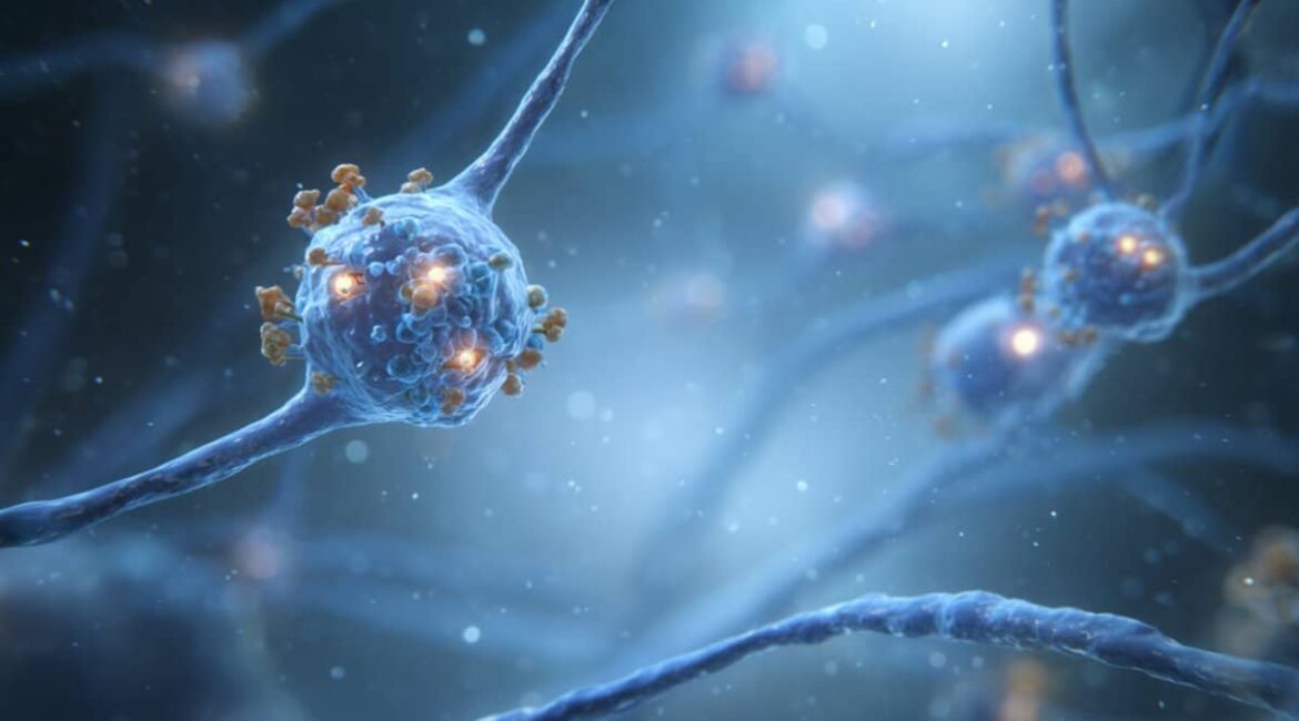
For the first time ever, researchers have used cryo-electron imaging to map the molecular structure of serotonin receptors in the brain. These receptors have a significant impact on how the cerebellum’s neurons speak, and they have an impact on movement, balance, and cognition.
Scientists hope to tell potential solutions that may recover work after injury or biological disruption by visualizing the receptor bound to proteins at synapses. This fundamental finding provides a roadmap for recovering broken head wires in motor and mental disorders, despite not being promptly applicable to treatments.
Important Information:
- Cryo-EM’s first-ever visualization revealed the cerebellar serotonin receptors ‘ architecture at near-atomic quality.
- Neural Precision: The receptors are arranged with precise spatial perfection when detecting neurotransmitter signals.
- Medical Potential: The findings provide the framework for synapse-targeting treatments to treat movement and cognition-related disorders.
Oregon Health and Science University as a cause
For the first time, researchers have studied the shape and function of important receptors connecting neurons in the body’s cerebellum, which is located behind the brain and is crucial for functions like coordination of movement, stability, and cognition.
The study, which was published today in the journal , Nature, provides new information that may lead to the development of treatments to fix these buildings when they are damaged by damage or genetic variants affecting motor skills like sitting, standing, walking, running, and jumping, as well as learning and memory.
Basic science research that won’t lead to a new drug or treatment, as demonstrated by scientists at Oregon Health & Science University, is the subject of a discovery that has been made by American researchers over the years to advance human health.
The National Institutes of Health and the Howard Hughes Medical Institute supported the new research, which was published in one of the most prestigious scientific journals in the world.
The study reveals the organization of a particular type of glutamate receptor, which is considered to be the brain’s primary excitatory neurotransmitter, bound together with proteins clustered on synapses, or junctions, between neurons in the cerebellum. This is a particular type of glutamate receptor.
” Synapses are crucial in all aspects of brain function, but how those components come together in a functional synapse,” said senior author and PhD, Eric Gouaux, Ph. senior scientist with the , OHSU Vollum Institute.
” Really important to have receptors organized in the right way so that they can detect neurotransmitters released by an adjacent cell.”
Glutamate in the cerebellum: an examination
Researchers examined the shape of a particular kind of glutamate receptor in the cerebellum of rodents at a near-atomic scale using OHSU’s state-of-the-art cryo-electron microscopy, which was established as one of three national centers in 2018 and housed in a reinforced basement of a building on the university’s South Waterfront Campus.
According to co-author Laurence Trussell, Ph. D.,” We know if there is an injury or genetic mutation in the cerebellum, it can lead to devastating disorders of balance, movement, or cognition.” Dr., Vollum Institute scientist and professor of otolaryngology/head and neck surgery at OHSU School of Medicine.
This particular glutamate receptor, according to the author, appears to be very important in how the cerebellum functions. It’s entirely possible that improving the function of these receptors by creating drugs that target them.
The Jennifer and Bernard Lacroute Endowed Chair in Neuroscience Research at OHSU and Gouaux, an investigator with the Howard Hughes Medical Institute, said the new discovery might have applications for novel treatments.
” We’ve been interested in this question about synapse engineering and molecular insight that may one day assist in repair damaged synapses,” he said. This has some promising new directions and potential therapeutic applications.
Chengli Fang, Ph. D., the lead author. Nearly all of the experiments reported in the publication were carried out by D., a postdoctoral researcher in the Gouaux lab.
Co-authors include , Cathy J. Spangler, Ph. D., and Trussell and Fang. D.,  , Jumi Park, Ph. OHSU, and OHSU’s Natalie Sheldon, both of OHSU and the Howard Hughes Medical Institute.
Funding: The National Institutes of Health, along with the National Cancer Institute, the National Institute of Neurological Disorders and Stroke, the National Institute of Deafness and Other Communication Disorders, all have funding for the research that was reported in this publication under the grant numbers K00CA253730, R01NS038631, R35NS116798, and R01DC004450.
The authors are solely responsible for the content, which does not necessarily reflect the NIH’s official opinion.
About this news about neuroscience research
Author: Erik Robinson
Source: Oregon Health and Science University
Contact: Erik Robinson – Oregon Health and Science University
Image: The image is credited to Neuroscience News
Original Research: Closed Access.
Eric Gouaux and colleagues ‘” Localization and noelin clustering of AMPA receptors that are natively Ca2+-permeable.” Nature
Abstract
Localization and noelin clustering of AMPA receptors that are natively Ca2+-permeable
The fast excitatory synaptic transmission is made possible by AMPARs, which are essential for motor coordination, learning, memory, and synaptic plasticity.
Although extensive structural studies have been done on recombinant AMPARs and native calcium impermeable ( CI) -AMPARs along with their auxiliary proteins, the molecular structure of native calcium permeable ( CP ) -AMPARs has remained a mystery.
We present the first visualization of these receptors, immunoaffinity purified from rat cerebella, and cryo-EM to reveal their subunit composition, physiological architecture, and gating mechanisms.
Our results show that the majority of assemblies are composed of GluA1 and GluA4, with the GluA4 subunit occupying the positions B and D, while auxiliary subunits, such as TARPs, are located at the B and D positions and CNIH or TARPs at the A and C positions, respectively.
Additionally, we improved the structure of the Noelin 1-GluA1/A4 complex, where Noelin 1 ( Noe 1 ) specifically binds to the GluA4 subunit at the positions B and D. Notably, Noe 1 stabilizes the amino-terminal domain ( ATD ) layer without affecting receptor gating characteristics.
Noe 1 supports AMPAR function by creating dimeric-AMPAR assemblies that are likely to be involved in extracellular networks, clustering receptors within synaptic environments, and modulating receptor responsiveness to synaptic inputs.
