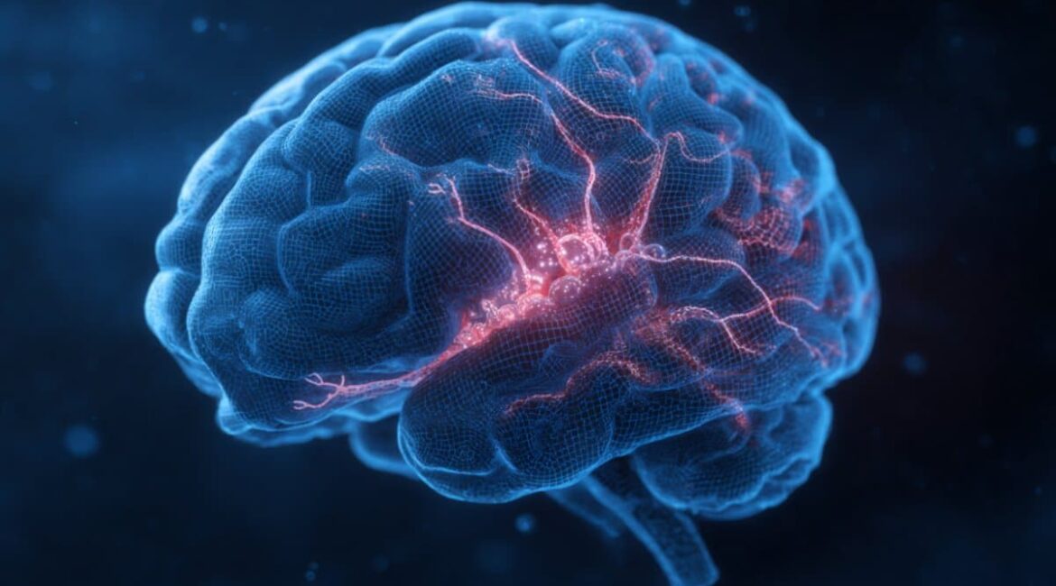
Summary: Researchers have discovered how the loss of the autism-linked protein PTEN in a particular set of antagonistic neurons reshapes brain circuits linked to fear and anxiety. They discovered that deleting PTEN from somatostatin-expressing brain neurons increased local suppression by 50 % while boosting activating input from nearby brain regions using advanced circuit-mapping techniques.
Without affecting cultural or repetitive behaviors that are frequently found in adhd, this imbalance caused animal designs to model higher levels of fear and anxiety. One of the most accurate depictions of how microcircuit changes may be responsible for various ASD-related traits is provided by the study, opening up new avenues for targeted treatments.
Important Information:
- Circuit-Specific Change: Somatostatin neurons lose PTEN and weaken nearby inhibition while amplifying key amygdala excitatory signals.
- Behavioral Outcome: The improved wiring was not related to any social or repetitive actions changes, but rather to increased fear and anxiety.
- Precision Mapping: Experts used a high-resolution implantable method to track microcircuit changes linked to genetic abnormalities.
Origin: Max Planck, Florida
Researchers at the Max Planck Florida Institute for Neuroscience have discovered how a gene that is implicated in autism and macrocephaly ( large head size ) rewires circuits and alters behavior after loss.
Their results, which were published in Frontiers in Cellular Neuroscience, reveal certain circuit changes in the brain brought on by PTEN decline in antagonistic neurons, giving new insights into the underlying circuit changes that contribute to heightened fear and anxiety.
One of the most significant dementia risk chromosomes has recently been identified as PTEN. This protein has variants in a sizable percentage of those who also have mind vegetation, making it a key component in the study of differences in brain function.
Researchers have used animal models to evaluate the effects of PTEN misregulation, where a global lowering in PTEN causes altered friendliness, repetitive behaviors, and increased anxiety, which are frequently linked to ASD in humans.
Animal designs that undermine PTEN throughout the nervous system have been challenging in terms of understanding how PTEN function leads to particular circuit and behavioral modifications.
So, Dr. McLean Bolton and her crew, head of the MPFI research team, have concentrated on the changes in the northern lateral brain caused by the loss of PTEN in somatostatin-expressing antagonistic neurons, a crucial cerebral people.
Through both human cells studies and biological mouse models, modifications in the function of antagonistic neurons have been observed in the development of ASD. Additionally, it is known that the PTEN gene controls the growth of antagonistic cells.
Thus, a cell-type-specific PTEN disruption in antagonistic neurons was a useful tool for studying specific circuit modifications that are related to ASD.
It is crucial to examine how genetic risk factors function within different neurological circuits, according to Dr. Bolton, even though a cell-type certain disruption does not recreate the genome-wide changes seen in humans.
Understanding these methods is a necessary first step in the development of targeted treatments for a certain set of traits, such as severe anxiety.
The group, led by Dr. Tim Holford, combined a biological unit that simply affected somatostatin-containing antagonistic neurons with a novel circuit tracking technique that was developed in the facility.
This method accurately characterized the electrical responses of individual neurons to the sequential optogenetic activation of hundreds of nearby neurons, allowing for quick mapping of connectivity and strength using the precision of electrical recordings and the scale of imaging techniques.
We can use this powerful technique to track down genetic variation-related changes in local neuron strength and connectivity.
We were interested in learning how the disruption of PTEN signaling in a single cell type would alter how the brain processes information and contribute to the broad ASD phenotype, according to Dr. Holford.
The findings were surprising because the researchers concentrated on the circuits in the central amygdala ( CeL ), a brain region known to inhibit the upstream expression of fear responses.
Deleting PTEN specifically in somatostatin-containing interneurons caused a 50 % disruption to local inhibitory connectivity in the CeL and significantly reduced the strength of the remaining inhibitory connections.
A rise in the volume of excitatory inputs from the basolateral amygdala ( BLA ), a nearby brain region that relays emotionally relevant sensory information to the CeL, was a contrast to this decreased connectivity between inhibitory connections within the CeL.
This imbalance in neural signaling was not related to alterations in social behavior or repetitive behavior patterns that are frequently found in ASD, but rather to heightened anxiety and increased fear learning, according to behavioral analysis of the genetic model.
The findings provide one of the most detailed maps to date showing how local inhibitory networks in the amygdala are impacted by genetic variations linked to neurological disorders, as well as confirm that PTEN loss in this particular cell type is sufficient to cause specific ASD-like behaviors.
Importantly, the altered circuitry did not affect all social interactions, which suggested that microcircuit changes may be the cause of PTEN-related anxiety and fear.
By exposing the local circuitry that underlies particular traits, as Dr. Holford puts it, we hope to be able to distinguish the functions of particular microcircuits within the scope of neurological disorders, which may one day aid in the development of targeted treatments for particular cognitive and behavioral characteristics.
We intend to examine these circuits in future studies to determine whether these microcircuit alterations are convergent and explain why people express more fear and anxiety in different genetic profiles.
About this news about autism, autism, and research into anxiety
Author: Lesley Colgan
Source: Max Planck Florida
Contact: Lesley Colgan – Max Planck Florida
Image: The image is credited to Neuroscience News
Open access to original research.
McLean Bolton et al., “PTEN in somatostatin neurons is necessary for central amygdala’s inhibitory synaptic connectivity and regulates fear and anxiety..” Cellular neuroscientists at the forefront
Abstract
PTEN in somatostatin neurons is necessary for central amygdala’s inhibitory synaptic connectivity and regulates fear and anxiety.
Introduction: Up to 25 % of ASD patients with macrocephaly have PTEN mutations, and the phosphatase and tensin homolog, which is deleted from chromosome 10, is a negative regulator of the mTOR pathway.
As do various mouse models with conditional knockouts of PTEN, germline PTEN haploinsufficiency mice exhibit behavioral characteristics resembling ASD. A number of genetic mouse models and human tissue studies have suggested that the GABAergic interneurons ‘ dysfunction may contribute to the development of ASD, but the precise mechanisms are still undetermined.
PTEN serves as a study’s target because it controls the development of inhibitory neurons produced by the medial ganglionic eminence, promoting the maturation and survival of parvalbumin ( PV+ ) neurons in contrast to somatostatin ( SOM+ ) neurons.
Utilizing behavioral analysis, electrophysiology, and two-photon local circuit mapping, we investigate how PTEN regulates SOM+ neurons at the cellular and circuit levels in the central lateral amygdala ( CeL ), which governs the key ASD behavioral symptoms of social anxiety and altered emotional motivation for social engagement.
Results: We discovered that SOM+ neurons ‘ knockout of PTEN causes increased levels of fear and anxiety, as well as a decrease in CeL local circuit connectivity. In particular, this manipulation altered the distribution of local connections in a cell-type specific way and reduced the strength of connections between individual neurons. The basolateral amygdala ( BLA ) was enhanced in contrast to the deficit in local inhibitory connections within CeL and the excitatory drive from the main CeL input.
Discussion: This combined imbalance of enhanced excitation and decreased local inhibition likely contributes to the heightened fear learning and anxiety we observed in the PTEN-SOM-KO mice.
