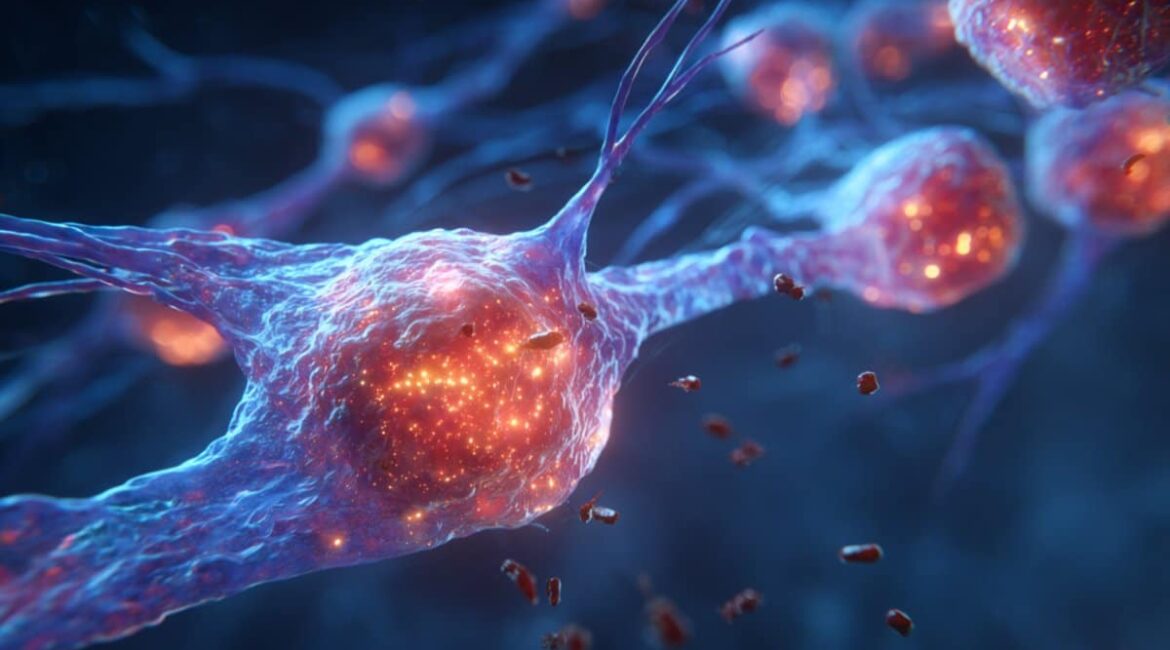
Generally speaking, the eye doesn’t conjure neutrophils, the body’s default first responders, when it is injured, unlike most tissues. Instead of requesting copy, microglia, the brain’s native immune cells, can deal with photoreceptor damage.
Researchers discovered this distinctive immune response in life mouse retinas using adaptive optics imaging. The findings point to a safe” cloaking” mechanism that stops excessive inflammation, which could help guide the development of new eye care options.
Important Information:
- Despite being close, neutrophils are unable to perform receptor maintenance.
- Just brain-resident immune cells can listen to ocular injury through microglia activation.
- Inflammation Shield: To stop the immune system from regenerating, the eye does inhibit recruitment.
University of Rochester
Neutrophil, an immune system found in the blood, are typically the first line of defense during the majority of eye diseases or injury.
However, researchers at the University of Rochester’s Del Monte Institute for Neuroscience and the Flaum Eye Institute have discovered that the eye reacts differently to many other body parts.
When receptor cells in the retina are damaged, microglia or the mind’s immune cells react and neutrophils are never recruited to assist despite passing through local blood vessels.
Jesse Schallek, PhD, associate professor of obstetrics and senior author of a study released today in , covering, said,” This getting has significant implications for what millions of Americans suffer vision loss as a result of photoreceptor loss.”
” This association between two important immune cell groups is essential information as we develop new solutions that require a thorough understanding of defense cell relationships.”
Researchers studied the insides of mice with receptor harm using adaptive optics imaging, a lens technologies developed by the University of Rochester that enables the scanning of individual neurons and immune cells inside the living eye.
They discovered that while both microglia and neutrophil cells are present in the retina, only microglia cells can heal from photoreceptor damage, and they do not use neutrophils to do so.
This, according to researchers, suggests a form of cloaking occurs during retinal injury to shield the retina from a rush of immune cells that could cause more harm than good.
The passing neutrophils are so close to the reactive microglia, and yet they do not signal to them to help with damage recovery, according to Schallek.
This is markedly different from what other body parts of the body, where neutrophils are the ones who initiate local damage and mount an early and robust response.
Photoreceptor cells are particular to the retina. Our brains, which are able to see, are then informed that light is being transformed into electrical and chemical signals.
There are currently no treatments for photoreceptor cells, including age-related macular degeneration, retinitis pigmentosa, and cone-rod dystrophy, which are all linked to a wide range of diseases.
This study has now demonstrated that it is possible to visualize the dynamics of individual cells as they communicate with one another and as the retina responds to damage.
The study was led by first author , Derek Power, a laboratory technician in the Schallek lab. Other authors include Genentech Inc.’s Justin Elstrott, PhD.
The National Eye Institute, Research to Prevent Blindness, the Dana Foundation, and Genentech, Inc., the funding for the study was provided by the National Eye Institute.
About this news about this research in visual neuroscience
Author: Kelsie Smith Hayduk
Source: University of Rochester
Contact: Kelsie Smith Hayduk – University of Rochester
Image: The image is credited to Neuroscience News
Open access to original research
” Despite strong microglial activation, photoreceptor loss does not recruit neutrophils.” by Derek Power et al. eLife
Abstract
Despite strong microglial activation, photoreceptor loss does not recruit neutrophils.
Tissue-resident immune cells like microglia and circulating systemic neutrophils are frequently the first ones to respond to a central nervous system ( CNS ) injury.
This is challenging for high-resolution imaging in vivo because of the low understanding of how these cells interact with CNS damage and even less so in the neural retina.
In this study, we use AOSLO ( fluorescence adaptive optics scanning light ophthalmoscopy ) to study neutrophils and microglia in mice.
We use label-free phase-contrast AOSLO at micron-level resolution to simultaneously monitor immune cell dynamics. 488 nm of light focused on photoreceptor ( PR ) outer segments was used to cause retinal lesions.
With minimal collateral damage to cells above and below the focus plane, these lesions focally ablated PRs.
We used in vivo AOSLO and OCT imaging to reveal the microglial and neutrophil response’s natural history from minutes to months after injury.
Despite being close to vessels that were only microns away from the vessel, neutrophils were not recruited despite a dynamic and progressive immune response with cells migrating into the injury locus within one day of injury. Post-mortem confocal microscopy confirmed in-vivo findings.
This research demonstrates that in response to acute, focal loss of PRs, a condition found in many retinal diseases, microglial activation does not recruit neutrophils.
