Answered: Major Issues
Q: Why do mental thoughts have a higher level of vividness than just negative people?
A: Because the brain and brain coordinate uniquely, which increases recall ability.
Q: What did the study reveal about how the mind functions while retrieving memories?
A: During both processing and retrieval, beta activity patterns created in the amygdala during mental processing were reactivated in the hippocampus.
Q: How does this finding affect how we understand PTSD and other problems?
A: It provides a possible mechanism for controlling aggressive emotional memories and provides a target for upcoming therapies that could alter or reverse maladaptive memory reactivation.
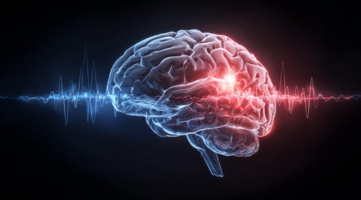
Summary: A new study examines how the brain and brain coordinate when forming and retrieving personal recollections from direct tapes from human neurons. High-frequency alpha activity in the brain influences cortical responses during aversive storage encoding, which are later reactivated in the hippocampus, but not the amygdala, during memory recall.
These patterns, which were excluded from negative memories, were primarily related to correctly remembered psychological scenes. The studies provide a molecular explanation for why emotionally charged experiences are frequently remembered more powerfully, and they also provide new treatment options for memory-related disorders.
Important Information:
- High beta bursts in the brain during personal events influence cortical activity.
- Hippocampus Stores Recurrence: During retrieval, the hippocampus merely after reactivates these amygdala-driven patterns.
- Possible Clinical Impact: Erkenntnisse gained from this study might lead to treatments for PTSD and anxiety disorders that goal memory recurrence.
Origin: Neuroscience News
Why do emotionally charged thoughts last so long in our minds while humdrum ones fade?
A new research utilizing cranial recordings of individual patients uncovers a fascinating neurological choreography between the brain and the hippocampus, which helps explain why aversive experiences frequently etch themselves deeply into storage.
The study, which was published in Nature Communications, reveals how specific patterns of brain activity are afterwards reactivated during storage retrieval, not in the amygdala, but in the hippocampus.
This discovery not only improves our understanding of personal recollection, but it may also provide insight into conditions like PTSD and anxiety.
Our brains don’t handle things emotionally charged like it would be treated in a normal situation, such as a traumatizing thought or a terrifying encounter. Instead, it employs professional frameworks to process and store that remembrance in a way that makes it easily recalled later.
The brain and the amygdala, both known for their roles in memory, work in concert to express these experiences. Scientists weren’t sure how this partnership translates into long-term memory development and subsequent retrieval up until now.
This study attempts to bridge that gap by demonstrating how the brain after uses the rhythmic high-frequency activity of the amygdala to imprint patterns.
Researchers observed the mind in action as participants watched psychological and natural scenes using elusive immediate recordings from epilepsy patients. Twenty-three patients had electrodes placed in their brain, and fourteen of them had ipsilateral hippocampus protection as well.
Over the course of two weeks, participants initially viewed scenes—some neutral, some aversive—and then made fundamental indoor/outdoor judgments. They were tested on their storage for these photos a day later, putting them in the categories of “remembered,” “known,” or “new.”
The outcomes were astounding. A certain increase in beta exercise (60–85 Hz ) in the hippocampus, which was triggered by accurately remembered aversive scenes, started about 0.7 seconds after the image was released. Ironically, the amygdala didn’t display this same retrieval-specific beta signature.
Rather, it appeared to be playing a role in shaping the memory at the time of processing. Hippocampal responses were found to be entrain during this period by high-frequency beta bursts (90–150 Hz ) in the brain. These designs were finally reactivated during both memory compression and subsequent recovery, but only in the hippocampus.
The researchers used a technique known as representational similarity analysis ( RS A ) to go deeper. This method made it possible to compare the patterns of brain activity observed during searching to those observed during processing.
Globally, the alpha action patterns during processing and retrieval were really decorrelated, suggesting that the brain isn’t just replaying the same signals.
However, when experts examined sharp, temporary spikes of activity known as rhythmic beta bursts in the amygdala, they discovered something astonishing: these accurate patterns were reactivated in the hippocampus.
Also more revealing was that the brain didn’t really silently echoe the brain. The synaptic activity mirrored the amygdala’s rhythmic gamma bursts with a postpone of about 0. 5 to 1 second during the initial encoding.
The brain after reactivated those exact certain patterns without the amygdala having to “remind” it. In fact, the hippocampus acted as a piece of music that the brain would later play solo.
Many controls were used to test the specificity of this recurrence. For instance, when researchers examined additional areas of the medial temporal cortex, they found no like pattern of recurrence.
Additionally, strange moment windows that are related to amygdala peaks did not demonstrate this phenomenon, which suggests that the recurrence is both framework- and time-specific.
The effects of this function are significant. Personal memories are frequently relived with distressing clarity in disorders like PTSD. Understanding the historical and fundamental dynamics of how memories are preserved and reactivated could lead to the development of new treatments to halt or modify these memories.
According to the researchers, brain stimulation techniques like amygdala theta-burst stimulation may one day be modified to change or enhance particular memory traces in response to medical needs.
In summary, this study illuminates a basic feature of human thinking: how we retain information. The study provides a powerful neural explanation for why some situations become unforgettable by showing that the amygdala doesn’t just signal psychological importance but really imprints its signature on the hippocampus. It turns out that the brain is a powerful and very selective storyteller rather than just a quiet recorder of experience.
One point is becoming evident as we learn how memory functions at the millisecond-scale brain rhythmic level: the story of personal memory is not just about how we feel it in the brain but also about when and where we experience it.
About this reports about research into psychological memory and neuroscience
Author:  , Neuroscience News Communications
Source: Neuroscience News
Contact: Neuroscience News Communications – Neuroscience News
Image: The image is credited to Neuroscience News
Start access to original analysis
Manuela Costa and colleagues ‘” Human synaptic recurrence of amygdala encoding-related beta patterns during aversive storage retrieval.” Nature Communications
Abstract
During aversive storage recovery, animal hippocampal reactivation of amygdala-related beta patterns during aversive memory retrieval.
The brain and hippocampus must coordinate their actions for personal memories.
An brain theta-hippocampal alpha phase code is involved in the formation of aversive memories, according to individual intracranial recordings.
However, it is still unclear what methods were involved in the re-entry and storage of aversive encounters.
Here, we demonstrate that synaptic gamma activity increases for accurately remembered aversive scenes by immediately recording from the individual amygdala and hippocampus.
Crucially, both aversive processing and searching result in patterns of high-amplitude gamma activity in the hippocampus but not the amygdala.
During aversive recovery, the hippocampus reactivates cortical gamma patterns that are trial-specific and exhibit highest visual similarity with amygdala activity at encoding.
This recurrence process takes place in the presence of gamma activity, which is usually unrelated to encoding and retrieval.
So, cyclic hippocampal gamma responses mimic the recovery of aversive memories, with activity patterns that appear to be ingrained by the brain during encoding.
