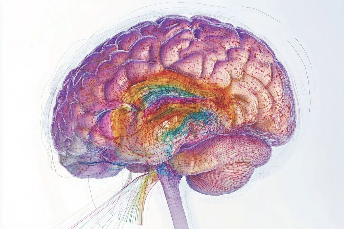
Summary: New research shows that not all brain cell time likewise, with certain cells, such as those in the brain, experiencing more age-related biological changes. These adjustments include increased immunity-related genes and decreased activity in cerebral hardware genes.
The findings provide a detailed image of age-sensitive head regions, providing insight into how aged might affect brain conditions like Alzheimer’s. This study was serve as a starting point for the development of treatments for aging-related brain changes and neurological conditions.
Important Information:
- Uneven Aging: Neuroendocrine cells and ventricle-lining tissues show the greatest age-related biological changes.
- Gene Activity Shifts: Aging reduces cerebral loop genes but rises immunity-related genes.
- Medical Possible: Modeling age-sensitive cells properly advise treatments for aging-related mind diseases.
Origin: NIH
Researchers from the National Institutes of Health ( NIH) have discovered that not all cell types in the brain age in the same way based on recent brain mapping research.
They found that some tissues, such as a small party of hormone-controlling tissues, may have more age-related changes in hereditary exercise than others.
The findings, published in , Nature, support the idea that some tissue are more vulnerable to the aging process and aging mental disorders than another.
” Aging is the most important risk factor for Alzheimer’s disease and many other catastrophic mental disorders. These findings provide a very detailed diagram for which brain tissue may be most affected by aging”, said Richard J. Hodes, M. D., chairman of NIH’s National Institute on Aging.
This new chart may ultimately affect how brain development is impacted by aging, as well as serve as a reference for the development of novel treatments for aging-related brain disorders.
Researchers used innovative genetic research tools to research individual cells in the hippocampus of 2-month-old “young” and 18-month-old “aged” animals.
Researchers analyzed the biological activity of a variety of cell types in 16 distinct large regions, which make up 35 % of the mouse brain’s overall volume. For each age, the study examined the activity of 3 different cell types.
The preliminary findings, like preceding studies, showed a decrease in the action of genes linked to cerebral circuits.
These decreases were observed in “glial” cells called astrocytes and oligodendrocytes, which is control serotonin levels and electric insulating nerve fibers. These are both seen in neurons, the main circuitry cells, and in “glial” cells called astrocytes and oligodendrocytes, which may help neurological signaling.
In comparison, aging increased the activity of chromosomes associated with the body’s resistance and aggressive systems, as well as mental blood vessel cells.
Further research revealed which body types may be the most susceptible to aging. For instance, according to the findings, aging lessens the number of newly developed cells present in at least three different mental regions.
Some of these kid neurons does play a role in the wiring that controls some forms of memory and learning, while others may aid mice in learning from distinct smells, according to recent research.
The second atrium, a major network that allows spinal liquids to pass through the brain, is where the organisms that appeared to be the most prone to aging reside.
Located at the base of the mouse head, the brain produces hormones that can control the body’s basic needs, including heat, heart rate, sleep, appetite, and appetite.
The results revealed that cells that line the third ventricle and neighboring hypothalamus neurons had the greatest changes in genetic activity as they grew older, including increases in immunity genes and decreases in genes linked to neuronal circuitry.
The authors noted that these observations are in line with earlier studies on a variety of animals that found connections between aging and body metabolism, including those that showed how intermittent fasting and other calorie-restricting diets can prolong life.
In particular, the ventricle-lining cells regulate the flow of nutrients and hormones between the brain and the body while the age-sensitive neurons in the hypothalamus are known to produce feeding and energy-controlling hormones.
More research is required to look into the biological mechanisms that underlie the findings and look for any potential connections to human health.  ,
The project was led by Kelly Jin, Ph. D., Bosiljka Tasic, Ph. D., and Hongkui Zeng, Ph. D., from the , Allen Institute for Brain Science, Seattle. The scientists used brain mapping tools — developed as part of the , NIH’s , Brain Research Through Advancing Innovative Neurotechnologies® , ( BRAIN ) Initiative , – , Cell Census Network ( BICCN)  , — to study more than 1.2 million brain cells, or about 1 % of total brain cells, from young and aged mice.
” For years, scientists have focused on one cell at a time, mostly on the effects of aging on the brain. Researchers can now study how aging affects a large portion of the brain using cutting-edge brain mapping tools made possible by the NIH BRAIN Initiative, according to John Ngai, Ph.D. D., director, The BRAIN Initiative®.
This study demonstrates that expanding our understanding of the human body can provide scientists with fresh insights into how the brain ages and how neurodegenerative diseases may interfere with normal aging processes.
Funding: This study was funded by NIH grants , R01AG066027 , and , U19MH114830.
About this news story about brain mapping and aging.
Author: Christopher Thomas
Source: NIH
Contact: Christopher Thomas – NIH
Image: The image is credited to Neuroscience News
Original Research: Open access.
” Transcriptomic signatures of healthy aging in mice are brain-wide, dependent on the type of cell.” by Kelly Jin et al. Nature
Abstract
Transcriptomic signatures of healthy aging in mice are brain-wide, dependent on the type of cell.
A gradual loss of homeostasis across various molecular and cellular functions can be characterized as biological ageing. There are thousands of different cell types in mammals ‘ brains, each of which may have a different level of age resilience or resistance.
Here we present a comprehensive single-cell RNA sequencing dataset containing roughly 1.2 million high-quality single-cell transcriptomes of brain cells from young adult and aged mice of both sexes, from regions spanning the forebrain, midbrain and hindbrain.
847 cell clusters are found in high-resolution clustering of all cells, which reveals at least 14 glial type-dominated clusters with age differences.
We identify 2, 449 distinct differentially expressed genes ( age-DE genes ) for a variety of neuronal and non-neuronal cell types at the broader cell subclass and supertype levels.
We observe common signatures with ageing across cell types, including a decline in the expression of genes related to neuronal structure and function in many neuron types, major astrocyte types and mature oligodendrocytes, and an increase in the expression of genes related to immune function, antigen presentation, inflammation, and cell motility in immune cell types and some vascular cell types.
Finally, we find that some of the cell types in the third ventricle of the hypothalamus, including tanycytes, ependymal cells, and some neuron types in the arcuate nucleus, dorsomedial nucleus, and paraventricular nucleus, express genes that are canonically related to energy homeostasis.
Many of these types exhibit both an increase in immune response and a decrease in neuronal function.
These findings point to the possibility that the mouse brain’s third ventricle serves as its ageing hub.
Overall, this study systematically outlines a dynamic landscape of cell-type-specific transcriptomic changes in the brain related to normal ageing. This will serve as a foundation for research into how ageing affects how well people age and how ageing and disease interact with one another.


