Summary: Researchers have discovered a special plant cell in the fresh head that can develop into a variety of different cell types, which might explain the link between autism and glioblastoma. These stem cells exhibit gene expression patterns that control first brain development, which may cause neurological conditions when disregarded.
The research links childish neurons that are active during brain growth with autism-related genes in a precise gene expression map. The findings opened strategies for targeting glioblastoma’s roots and better knowing autism’s development roots.
Essential Information
- Stem cell identification: A stem cell that can differentiate into three different types of brain cells may be the catalyst for glioblastoma development.
- Autism Insight: Autism-associated genes are effective during important levels of brain growth, potentially affecting cerebral progress.
- Modern Mapping: A detailed analysis of the gene expression of brain cells reveals connections between growth and illness.
Origin: UCSF
Researchers at UCSF have discovered a plant cell in the fresh head that can differentiate cells from those found in lesions. The discovery might shed light on how older brain cells exploit development mechanisms to promote the rapid growth that deadly brain cancers like glioblastoma do.
They made the discovery by conducting a thorough genetic analysis of human brain tissue from the first two decades of life. The conclusions appear Jan. 8 in , Nature.  ,
” Some head diseases begin during different stages of development, but until now we haven’t had a detailed blueprint for just understanding good mental growth”, said Arnold Kriegstein, MD, PhD, professor of neuroscience at UCSF and co-corresponding author of the paper.
Our map reveals the hereditary blueprints that underlie the development of the human brain and how these processes malfunction in response to particular types of brain dysfunction.
The study examined the expression of genes in tissue extracted from donated head samples. To aid in explaining how the brain makes connections, the experts kept track of the cells ‘ unique locations.  ,
The information also contained suggestions about autism’s origins, in addition to the discovery of an earlier stem cell that might explain the biology of glioblastoma in age. The experts have made the information available for use in the area to learn about a variety of other mental disorders.
” Our study paints one of the most thorough pictures of people head advancement”, said Li Wang, PhD, postdoctoral scholar in Kriegstein’s labs and co-first and co-corresponding author of the paper.
We’re excited to see what other fields can do with the painful information, and we’re excited to see how ideas based on observations in the doctor and laboratory can now be tested against it.
Tests reveal a gold trove ,
The majority of research into the developing brains is done using dog models, which are at best shed proxies for the human mind.  ,
The group, also led by co-first artist Cheng Wang, PhD, and co-corresponding artist Jingjing Li, PhD, wagered that valuable new insights may be made by studying the human mind itself. They worked with the National Institutes of Health ‘s , NeuroBioBank , and local hospitals associated with UCSF to obtain brain samples.
These samples, which were donated from 27 people from early life to adolescence, were sent to UCSF to be tested for gene expression in thousands of individual cells.  ,
Gene expression refers to how DNA, stored in chromosomes, is copied into RNA– short-lived genetic messages– which are then used as a template for building proteins. The researchers could examine the behavior of those cells by measuring RNA.
” RNA degrades quickly, and you need to have very pristine tissue in order to get usable data”, Kriegstein said.
We thank the community for funding such important research by donating this priceless tissue, and it was a huge breakthrough for Li and his colleagues to conduct such high-resolution genomic tests on this tissue.
The researchers determined which regions of each chromosome could be used to express genes in which cell. Additionally, they indicated where each cell had been removed from the brain.  ,
The researchers concentrated on cells taken from the front and the back of the cerebral cortex, areas that humans have in charge of language, memory, and learning.
” RNA alone doesn’t tell the entire story of a cell’s behavior”, Wang said.
We could begin to understand the full story of brain development by measuring the RNA and chromatin states at the same time in the same cell and then mapping each cell back into the brain’s structure.
In the developing brain, a mix of autism risk genes manifest itself.
Autism isn’t caused by a single gene mutation, but rather by the combination of many gene mutations.  ,
The researchers discovered that immature neurons had activated many of the genes responsible for autism before symptoms started to show. Mutations in these genes, they said, could interfere with the growth of the young brain, leading to autism.
When young neurons were still migrating throughout the growing brain and figuring out how to establish connections with other neurons, these gene expression programs became active, Wang said.
” Those maturing neurons might become perplexed about where to go or what to do if something goes wrong at this point.”
It’s still unclear exactly how autism develops in the brain because the study didn’t examine tissue from those with autism. However, the research links the cells that make up the growing brain to many genetic variations that are linked to autism.
In a sense, we’ve identified many of the dots driving autism at a critical point in development, according to Kriegstein.” People talk about connecting the dots to come up with a picture of how autism emerges.
” This area of development may merit further investigation to unravel all the autism mysteries.”
Could a program that starts with early brain growth be switched to later in life to stop tumor growth?
As the researchers compared their data, Wang observed a group of stem cells that appeared to be working on something unusual. These immature cells were beginning to express genes that were unique to three mature cell types.
Many stem cells in the developing brain are just one cell type, such as a neuron or support cell. Some people can have two types of maturation. However, these stem cells could also develop into three different lignins: glia, glia, and neuron, respectively.
The researchers speculated that this trait might help it develop glioblastoma tumors, which have three similar cell types, later in life.
” Glioblastoma has been challenging because it’s so heterogeneous”, Kriegstein said. ” Li discovered a precursor capable of producing all three glioblastoma cell types.”
The finding supports a well-known theory that tumors mislead genetic growth strategies to cause excessive growth in adulthood.  ,
And it might offer a fresh approach to treating” cancer stem cells,” the source of glioblastoma.
We might be able to stop that growth by understanding the context in which one stem cell produces three different types of cells in the developing brain when it reappears during cancer, Wang said.  ,
Authors , In addition to Wang, Wang, Li, and Kriegstein, other UCSF authors are Juan A. Moriano, PhD, Songcang Chen, MD, Guolong Zuo, PhD, Arantxa Cebrián-Silla, PhD, Shaobo Zhang, MD, Tanzila Mukhtar, PhD, Shaohui Wang, Mengyi Song, PhD, Lilian Gomes de Oliveira, MA, Qiuli Bi, Jonathan J. Augustin, PhD, Xinxin Ge, PhD, Mercedes F. Paredes, MD, PhD, Eric J. Huang, MD, PhD, Arturo Alvarez-Buylla, PhD, and Xin Duan, PhD.
Funding and disclosures:  , The work was supported in part by grants from the NIH ( U01MH114825, R35NS097305, P01NS083513, R01NS123912, K99MH131832 ). Kriegstein is a co-founder, consultant and director of Neurona Therapeutics. For all other funding, see the paper.
About this genetics, brain cancer, and autism research news
Author: Levi Gadye
Source: UCSF
Contact: Levi Gadye – UCSF
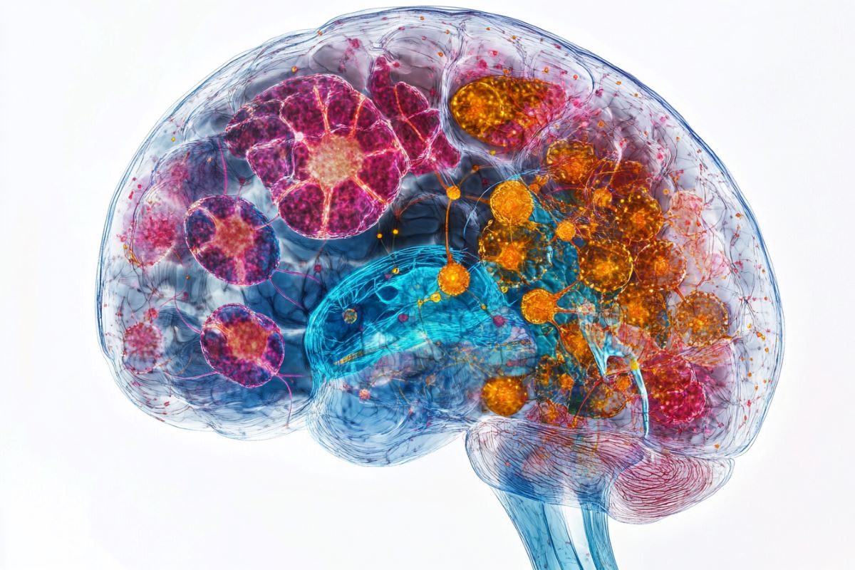
Image: The image is credited to Neuroscience News
Original Research: Open access.
Arnold Kriegstein and colleagues ‘” The developing human neocortex exhibits both cellular and molecular dynamics.” are the paper’s title. Nature
Abstract
The developing human neocortex exhibits both cellular and molecular dynamics.
The human neocortex develops in a highly dynamic manner, involving complex cellular pathways that are controlled by gene regulation.
We gathered paired single-nucleus chromatin accessibility and transcriptome data from 38 human neocortical samples that included both the primary visual cortex and the prefrontal cortex.
These samples span five main developmental stages, ranging from the first trimester to adolescence.
To demonstrate spatial organization and intercellular communication, we conducted spatial transcriptomic analysis on a subset of the samples in parallel.
This atlas enables us to catalogue cell-type-specific, age-specific and area-specific gene regulatory networks underlying neural differentiation.
Moreover, combining single-cell profiling, progenitor purification and lineage-tracing experiments, we have untangled the complex lineage relationships among progenitor subtypes during the neurogenesis-to-gliogenesis transition.
Tripotential intermediate progenitor cells ( Tri-IPCs ) are a tripotential intermediate progenitor subtype that is responsible for the local production of astrocytes, oligodendrocyte precursor cells, and astrocytes.
Notably, the majority of glioblastoma cells resemble Tri-IPCs at the transcriptomic level, which suggests that cancer cells participate in developmental mechanisms that promote growth and heterogeneity.
Furthermore, by integrating our atlas data with large-scale genome-wide association study data, we created a disease-risk map highlighting enriched risk associated with autism spectrum disorder in second-trimester intratelencephalic neurons.
Our investigation provides insight into the cellular and molecular dynamics of the developing human neocortex.


