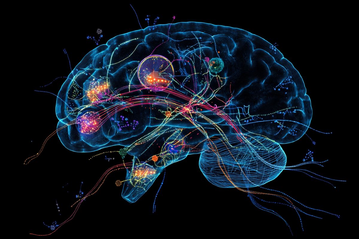
Summary: A comprehensive study mapped neuronal IL-1R1 ( nIL-1R1 ) expression in the mouse brain, highlighting its role in sensory processing, mood, and memory regulation. Researchers discovered that neurons that express IL-1R1 integrate both defensive and neural indicators, revealing connections between swelling and mental illnesses like depression and anxiety.
The research pinpointed important regions, such as the sensory cortex and brain, where IL-1 indicating influences synapse organization and neuronal circuit modulation. Importantly, cerebral IL-1R1 modifies neural pathways without triggering infection, suggesting specific features in the central nervous system.
Important Information:
- Immune-Neural Connection: Neuronal IL-1R1 integrates defensive signals into neurological circuits, influencing visual, disposition, and memory characteristics.
- Distinct Signaling Pathways: Unlike defense cells, cells with IL-1R1 regulate neural institution without causing irritation.
- Target Regions: IL-1R1 appearance is important in areas like the brain and visual cortex, linked to feeling rules and visual processing.
Origin: FAU
Interleukin-1 ( IL-1 ) is a key molecule involved in inflammation and plays an important role in both healthy and diseased states.
In condition, higher levels of IL-1 in the mind are linked to neuroinflammation, which can disrupt the body’s pressure response, cause sickness-like behaviors, increase inflammation by activating brain immune cells, and help immune cells from the body to enter the brain. Support cells can also produce harmful molecules, which could cause brain damage.
Elevated IL-1 levels are associated with mood disorders, such as depression, and problems with memory and thinking.
Conversely, in normal conditions without inflammation, IL-1 has essential roles in the brain. It helps regulate hormone activity, supports healthy sleep patterns, and improves cognitive functions such as memory and learning.
IL-1R1 is like a doorbell on cells that gets rung when there’s an infection or injury, and in immune cells, it signals the body to start an immune response.
However, neurons that express IL-1R1 are not thought to induce inflammation, suggesting that these cells may actually integrate immune signals into neural ones. It has yet to be revealed where or how IL-1R1 ( Interleukin-1 Receptor Type 1 ) may control or modify normal brain function.
Now, a new study by , Florida Atlantic University , provides the most detailed and comprehensive mapping of neuronal IL-1R1 ( nIL-1R1 ) expression in the mouse brain to date, resolving long-standing inconsistencies.
Previous research has suggested that IL-1 signaling in neurons is involved in sickness behaviors, anxiety, and changes in sleep, but the exact neural circuits involved have not been well-defined.
The study, published in the , Journal of Neuroinflammation, narrows down the specific neuronal populations and neurotransmitter systems that could mediate these effects.
Utilizing a clever cell tagging technique, researchers were able to identify neuronal populations that expressed nIL-1R1, giving new insights into the central nervous system’s ( CNS ) functional roles.
Previous studies conducted by the FAU Quan Laboratory, reveal that chronic IL-1 signaling in glutamatergic neurons influences cognitive and social-avoidance behaviors, particularly in the context of neuroinflammation and stress-related disorders.
This supports the hypothesis that the unique neural circuits described in this study, including those caused by chronic stress, depression, and anxiety, could be crucially affected by nIL-1R1.
Using genetically modified mice, researchers identified neurons in certain brain areas such as the , somatosensory cortex, hippocampus and others, which have neuronal IL-1R1. The majority of these neurons use glutamate, a neurotransmitter, for signaling, while others use serotonin, a crucial mood-related neurotransmitter.
They found that these IL-1R1-positive neurons are involved in circuits that control sensory processing, mood regulation and memory.
” Our study shows how certain neurons are connected to immune signals and may help explain how inflammation contributes to sensory, mood and memory disorders”, said , Ning Quan, Ph. D., senior author, professor of biomedical science, FAU , Schmidt College of Medicine, and an investigator in the FAU , Stiles-Nicholson Brain Institute.
These conclusions may provide new treatments for brain disorders linked to inflammation. In terms of behavioral implications, our results support the hypothesis that nIL-1R1 signaling influences emotional and cognitive behavior”.
Results show that nIL-1R1 expression is most prevalent in the somatosensory and glutamatergic systems, which were previously neglected in this regard.
Among the brain regions identified as expressing nIL-1R1, the dentate gyrus ( DG) was consistently highlighted, reaffirming its role as a key site for neuronal IL-1R1 expression.
The study also provides a detailed description of the thalamic relay centers and various sensory cortical regions, which suggests that IL-1 signaling might be important for sensory processing.
According to Dan Nemeth, Ph. D.,” This new discovery raises questions about whether immune signals influence our sensory processing and whether IL-1R1-mediated alterations of sensory signals contribute to cognitive issues, anxiety, or depression.” D., first author and a postdoctoral fellow, FAU Schmidt College of Medicine and Stiles-Nicholson Brain Institute.
” Furthermore, this study shows that neurons do not signal the same way other IL-1R1-expressing cells do”.
While researchers found neuronal IL-1R1 in brain regions related to mood, affect and cognition, an unexpected finding is that IL-1R1 is expressed in neurons in the sensory system.
Using high-tech spatial transcriptomics, they identified that neuronal IL-1R1 regulates gene pathways involved in synapse organization without triggering typical inflammation. This suggests that synaptic formation and function can be altered by IL-1R1.
” With the most detailed mapping of neuronal IL-1R1 expression in the mouse brain to date, this study brings an unprecedented level of clarity to how IL-1 signaling impacts the neural circuits that govern behavior”, said , Randy D. Blakely, Ph. D., co-author, executive director of the , FAU Stiles-Nicholson Brain Institute, the David J. S. Nicholson Distinguished Professor in Neuroscience, and a professor of biomedical science in FAU’s , Schmidt College of Medicine.
The findings provide crucial insights into the mechanisms underlying both normal and abnormal behavioral states seen in stress-related disorders, depression, and anxiety, according to the authors.
About this news from neuroscience research
Author: Gisele Galoustian
Source: FAU
Contact: Gisele Galoustian – FAU
Image: The image is credited to Neuroscience News
Original Research: Open access.
Ning Quan et al.,” Localization of brain neuronal IL-1R1 reveals specific neural circuits responsive to immune signaling.” Journal of Neuroinflammation
Abstract
Localization of brain-neuronal IL-1R1 reveals specific neural circuits that are immunely receptive.
Interleukin-1 ( IL-1 ) is a pro-inflammatory cytokine that is linked to the etiology of affective and cognitive disorders and has a wide range of neurological and immunological effects throughout the central nervous system ( CNS ).
The cognate receptor for IL-1, Interleukin-1 Receptor Type 1 ( IL-1R1 ), is primarily expressed on non-neuronal cells ( e. g., endothelial cells, choroidal cells, ventricular ependymal cells, astrocytes, etc. ) throughout the brain. However, the presence and distribution of neuronal IL-1R1 ( nIL-1R1 ) has been controversial.
For the first time, a novel genetic mouse line ( Il1r1GR/GR ) was used to map all brain nuclei and identify the neurotransmitter systems that express nIL-1R1 in adult male mice. It is a new genetic mouse line that allows for the visualization of IL-1R1 mRNA and protein expression.
In vivo, the direct responsiveness of nIL-1R1-expressing neurons to both inflammatory and physiological levels of IL-1 were evaluated.
In discrete glutamatergic and serotonergic neuronal populations in the somatosensory cortex, piriform cortex, dentate gyrus, and dorsal raphe nucleus, discrete glutamatergic and serotonergic neuronal populations were found.
Glutamatergic nIL-1R1 comprises most of the nIL-1R1 expression and, using , Vglut2-Cre-Il1r1r/r , mice, which restrict IL-1R1 expression to only glutamatergic neurons, an atlas of glutamatergic nIL-1R1 expression across the brain was generated.
Analysis of functional outputs of these nIL-1R1-expressing nuclei, in both , Il1r1GR/GR , and , Vglut2-Cre-Il1r1r/r , mice, reveals IL-1R1+ , nuclei primarily relate to sensory detection, processing, and relay pathways, mood regulation, and spatial/cognitive processing centers.
Intracerebroventricular ( i. c. v. ) injections of IL-1 ( 20 ng ) induces NFκB signaling in IL-1R1+ , non-neuronal cells but not in IL-1R1+ , neurons, and in , Vglut2-Cre-Il1r1r/r , mice IL-1 did not change gene expression in the dentate gyrus of the hippocampus ( DG).
GO pathway analysis of spatial RNA sequencing 1mo after restoration of nIL-1R1 in the DG neurons revealed that IL-1R1 expression downregulates genes that are involved in both synaptic function and mRNA binding while raising the levels of some complement markers ( C1ra and C1qb ).
Further, DG neurons exclusively express an alternatively spliced IL-1R Accessory protein isoform ( IL-1RAcPb), a known synaptic adhesion molecule.
Altogether, this study reveals a unique network of neurons that can respond directly to IL-1 via nIL-1R1 through non-autonomous transcriptional pathways, earmarking these circuits as potential neural substrates for immune signaling-triggered sensory, affective, and cognitive disorders.


