
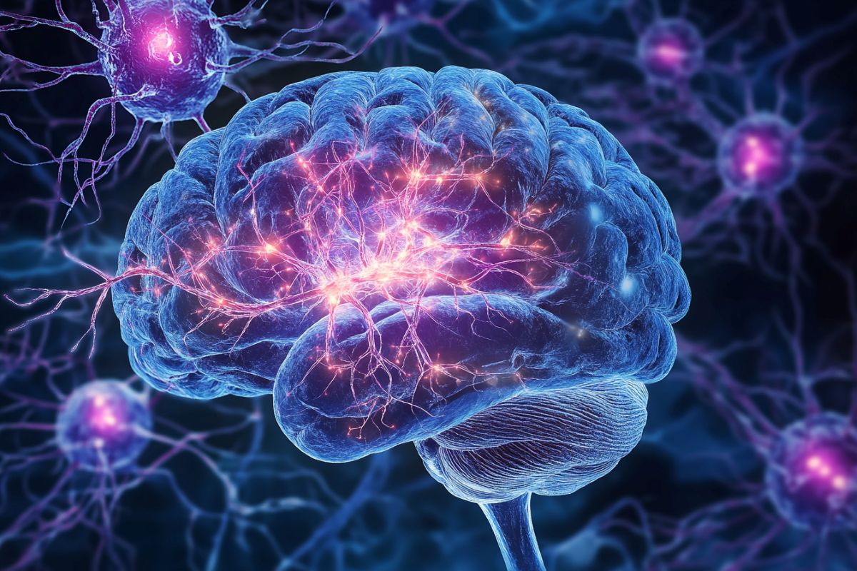
Summary: Researchers have identified memory-related brain tissue that are very susceptible to protein protein concentration, a key element in Alzheimer’s disease. Using the MISS brain-mapping approach, they profiled 1.3 million cells in animals to define which body types are most impacted.
Their studies show that glutamatergic neurons in the hippocampus are particularly vulnerable, while cerebral cells and neurones appear more resilient. Amazingly, the supply of brains cell types predicted tau formation better than genetic risk factors.
This suggests that biological composition, rather than genetics only, plays a vital part in Alzheimer’s development. The investigation offers useful insight that may guide coming work to slow or prevent the disease.
Important Information:
- Cell Type Vulnerability: Glutamatergic neurons in the hippocampus are more susceptible to protein formation.
- Safe Brain Cells: Oligodendrocytes appear less disturbed, possibly playing a defensive position.
- Beyond Genetics: The supply of brains body types predicts beta formation stronger than genetic risk factors.
Origin: UT Arlington
Researchers from The University of Texas at Arlington and the University of California–San Francisco have used a fresh brain-mapping technique to identify memory-related brain cells prone to proteins buildup, a crucial factor in the development of Alzheimer’s disease, an chronic, progressive brain disorder that gradually destroys memory and thinking skills.
In Texas, nearly half a million people live with Alzheimer’s disease, a form of dementia that costs the state approximately$ 24 billion in caregiver time, according to the , Texas Department of State Health Services.
Texas ranks fourth in the nation for Alzheimer’s cases and second in Alzheimer ‘s-related deaths.
To understand why certain parts of the brain are more affected by Alzheimer’s disease, researchers focused on tau, a protein that accumulates in brain cells and disrupt normal activity.
Using the MISS ( Matrix Inversion and Subset Selection ) mapping technique, which profiled approximately 1.3 million cells, the research team created detailed maps of different cell types in the brains of mice. They compared these maps to areas where tau builds up to identify which cell types are most affected.
Their findings are published in the peer-reviewed journal , Nature Communications Biology.
” Using mathematical and computational models, we found that certain cells in the hippocampus, a brain area important for memory and navigation, are more vulnerable to tau buildup”, said author Pedro Maia, an assistant professor of mathematics at UTA.
” These glutamatergic neurons showed a strong connection with tau deposits, meaning they are more likely to be affected. In contrast, brain cells in the cortex—the part of the brain that controls movement, sensory information, emotions and reasoning—were less likely to be affected by tau”.
Interestingly, the researchers also discovered that oligodendrocytes, brain cells that help insulate nerve fibers, were less affected by tau. This suggests that these cells might help to protect the brain against tau buildup.
The study also found that the distribution of different cell types in the brain may better predict where tau accumulation occurs than genetic factors alone. This implies that the types of cells present in different brain regions may be more important than Alzheimer ‘s-related genes in determining vulnerability to tau.
” Overall, this study helps us understand why certain brain regions are more affected by tau buildup leading to Alzheimer’s disease”, Dr. Maia said.
” By identifying the cell types and gene functions involved, our study showcases how theoretical and computational models can provide new insights into the progression of Alzheimer’s disease.
” This is another piece of valuable data that will help us specifically target the vulnerable cells and genes associated with tau buildup, potentially slowing or preventing Alzheimer’s disease progression in the future.”
To learn more about Maia’s work, visit the , NSF RTG Training in Mathematics for Human Health , program, which he co-leads on computational neurology.
Funding: This work was supported by the following NIH grants: R01NS092802, RF1AG062196 and R01AG072753.
About this brain mapping and Alzhiemer’s disease research news
Author: Katherine Bennett
Source: UT Arlington
Contact: Katherine Bennett – UT Arlington
Image: The image is credited to Neuroscience News
Original Research: Open access.
” Searching for the cellular underpinnings of the selective vulnerability to tauopathic insults in Alzheimer’s disease” by Pedro Maia et al. Communications Biology
Abstract
Searching for the cellular underpinnings of the selective vulnerability to tauopathic insults in Alzheimer’s disease
Neurodegenerative diseases such as Alzheimer’s disease exhibit pathological changes in the brain that proceed in a stereotyped and regionally specific fashion. However, the cellular underpinnings of regional vulnerability are poorly understood, in part because whole-brain maps of a comprehensive collection of cell types have been inaccessible.
Here, we deployed a recent cell-type mapping pipeline, Matrix Inversion and Subset Selection ( MISS), to determine the brain-wide distributions of pan-hippocampal and neocortical cells in the mouse, and then used these maps to identify general principles of cell-type-based selective vulnerability in PS19 mouse models.
We found that hippocampal glutamatergic neurons as a whole were significantly positively associated with regional tau deposition, suggesting vulnerability, while cortical glutamatergic and GABAergic neurons were negatively associated.
We also identified oligodendrocytes as the single-most strongly negatively associated cell type. Further, cell-type distributions were more predictive of end-time-point tau pathology than AD-risk-gene expression.
Using gene ontology analysis, we found that the genes that are directly correlated to tau pathology are functionally distinct from those that constitutively embody the vulnerable cells.
In short, we have elucidated cell-type correlates of tau deposition across mouse models of tauopathy, advancing our understanding of selective cellular vulnerability at a whole-brain level.