
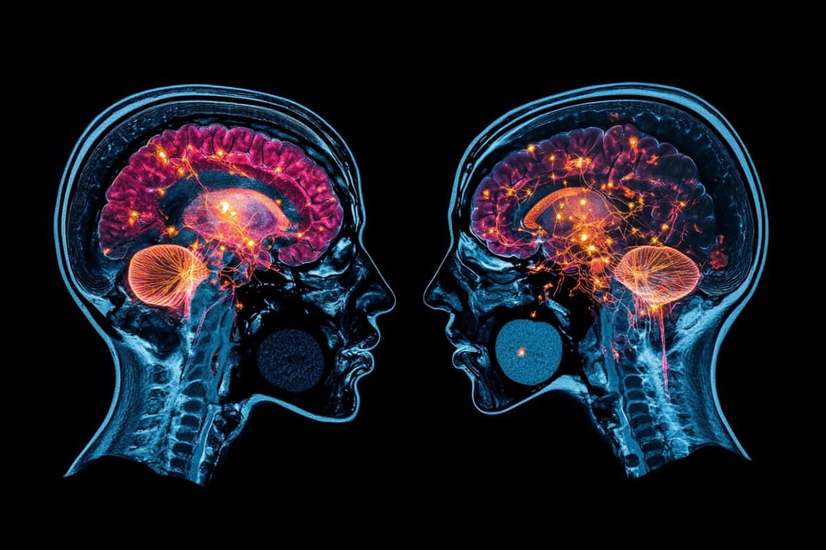
Summary: New research has revealed that treatment can cause breast cancer patients ‘ mind connection to change rapidly and widely. Researchers found irregularities in the frontal-limbic structure and cerebral cortex, which are both involved in professional function and memory, using functional MRI imaging.
As the treatment progressed, these shifts increased and intensified, suggesting that brain work had been altered by all at once. The findings may help explain the mental issues that are frequently encountered during and after chemotherapy, more commonly known as” treatment mind,” in the head.
Important Information:
- Rapid Brain Changes: Chemotherapy immediately alters brain communication.
- Affected Regions: Problems were discovered in areas that are related to memory and decision-making.
- Liberal Effect: Over time, accessibility changes have spread and worsened.
Origin: Wiley
Recent research published in the journal of Magnetic Resonance Imaging  has found changes in brain communication during treatment in breast cancer patients.
In a study of 55 breast cancer patients and 38 control brain cancer patients, researchers performed functional magnetic resonance imaging scans of the respondents ‘ brains over the course of several months.
Throughout the course of treatment, scans from patients showed changes in brain connectivity, particularly in the frontal-limbic system ( involved in executive functions ) and the cerebellar cortex ( linked to memory ).
As treatment went on, these changes worsened and spread.
The findings “imply that chemotherapy is rapidly disrupt brain work in breast cancer patients, potentially causing mental problems,” the authors wrote.
About this study in neuroscience and chemotherapy
Publisher: Sara Henning-Stout
Source: Wiley
Contact: Sara Henning-Stout – Wiley
Image: The image is credited to Neuroscience News
Original Research: Disclosed exposure.
Jing Yang and colleagues ‘” Improved brain practical networks in patients with breast cancer after various neoadjuvant chemotherapy cycles” Journal of Magnetic Resonance Imaging
Abstract
altered mind functions in breast cancer patients following various neoadjuvant treatment cycles
Background
Breast cancer (BC ) patients’ quality of life after chemotherapy is affected by cancer-related cognitive impairment ( CRCI). Although recent studies have looked at its neurological correlates, one time-point designs cannot account for how these changes change over time.
Purpose
To examine changes to the brain connectome of BC patients over a number of time points during neoadjuvant chemotherapy ( NAC ).
Study Method
Longitudinal.
Subjects
At baseline ( TP1 ), the first cycle of NAC ( TP2, 30 days later ), and the end ( TP3, 140 days later ), 55 BC patients underwent clinical assessments and fMRI. The assessments were conducted in two matched female healthy control ( HCs,  , n = 20 and n = 18 ).
Field Strength/Sequence
3D T1-weighted magnetization-prepared rapid gradient echo sequence at 3.0 T and rs-fMRI ( gradient-echo EPI).
Assessment
Graph concept techniques were used to analyze brain useful networks. We compared changes in brain international metrics to clinical scales ( including mood and cognitive tests ) and found that these changes were related to each other.
Patients were classified into subgroups based on their medical history, treatment regimen, and menopausal status. For each group, vertical analysis was performed at three different time points.
Statistical evaluations
An independent sample t–test for TP1 patient-HC evaluation paired  research and a t–test for vertical changes. Analysis examination of the connections between changes in network measurements and changes in clinical symptoms. Impact was defined as , p <, 0.05.
Results
Post-NAC, BC patients showed increased international performance ( TP2-TP1 = 0.087, TP3-TP1 = 0.078 ), decreased quality path length ( TP2-TP1 = −0.413, TP3-TP1 = −0.312 ), and altered lateral centralities primarily in the frontal-limbic system and cerebellar cortex. These abnormalities increased significantly as the chemotherapy progressed ( TP2 vs. TP3 ). T he T
The changes in opological criteria were also substantially related to those in the scientific scales. No variations were found within or between Hca groups ( p = 0.490–0.989 ) or BC groups ( p = 0.053–0.988 ) at TP1.
Opinions of the files
At TP2, NAC has an impact on the brain useful electric of BC patients, and these changes continue to grow and grow even more so at TP3.
Information Level
2.
Technical Performance
Stage 5.