
Summary: Researchers have created a biological “barcoding” technique to track the development of embryonic stem cells into cells and inner ear tissues. The team followed the development of premature stem cells and mapped the inner ear’s formation by labeling them in mice.
They discovered that two major stem cell types are responsible for the development of hearing-critical cell, giving researchers hints for upcoming hearing loss treatments. This method makes sense of the nervous system’s growth in a comprehensive “family trees,” which may ultimately lessen the need for pet testing.
Important Information:
- Biological Barcoding: A virus-based id made it possible for researchers to track stem cell growth into neurons and inner hearing cells.
- Hearing Insight: Essential inward ear structures are created by two primary plant cell types.
- Potential applications: The approach may aid in the study of other nervous system components and aid in hearing loss treatments.
Origin: Karolinska Institute
Analysts at Karolinska Institutet have created a method to demonstrate how an embryo’s nervous system and sensory organs are created.
By identifying stem cells with a biological “barcode,” researchers were able to study the development of the cell and learn how the inner ear developed in animals.
The finding, which was made in Science, might have important insight for the treatment of hearing loss in the future.
Our research illustrates how various cell types can be created from embryonic stem cells and how they are organized to form crucial mental structures, says docent at the Karolinska Institutet’s Department of Cell and Molecular Biology.
” We have created a family tree for the inner ear and nervous system cell,” one might say.
repairing damaged tissue
The analysts employed a method that involved injecting a pathogen into keyboard stem cells at a young stage of development. The virus contained a genetic “barcode” that was incorporated into the stem cells ‘ DNA and therefore passed down as the cell divided.
The researchers may observe the development of the cells into various neuronal and inner ear cell types by following this script.
The results demonstrated that two main types of stem cells are used to create cells in the inner ear, which are essential for reading. This information may lead to novel therapies for hearing damage.
Emma Andersson notes that studying the mechanisms that underlie hearing loss “uniquely gives us an opportunity to understand the fundamental mechanisms behind hearing lost.” It can lead to the development of novel methods for replacing or repairing damaged inward ear cells.
Exploring the nerve structure
The staff intends to use the technique to research both how the body’s internal organs develop and how different parts of the nervous system function. They hope that several genetic and developmental diseases may be treated and improved with their work.
Emma Andersson says,” We are only at the beginning of understanding the intricate mechanisms behind nervous system development.”
” Our approach opens up many fascinating new ways to look at how the nervous system and the rest of the body are created during fetal development. Additionally, the method may reduce the number of rabbits employed in research.
She co-led the investigation with postdoctoral fellow Jingyan He and former PhD student Sandra de Haan of Emma Andersson’s research group.
Funding: The study was supported by the Karolinska Institutet, the European Union, Horizon Europe, the Erling-Persson Foundation, the Swedish Research Council, the Knut and Alice Wallenberg Foundation, the Hearing Research Fund, and Wallenberg Bioinformatics Support. Jonas Frisén, a co-author, works for 10x Genomics as a specialist.
No additional conflicts of interest are disclosed.
About this information from biology and neurodevelopment study
Publisher: Emma Andersson
Source: Karolinska Institute
Contact: Emma Andersson – Karolinska Institute
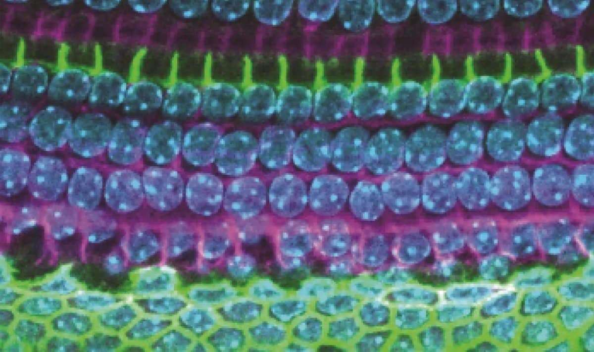
Image: The image is credited to Sandra de Haan
Initial research has been made private.
Emma Andersson et al., “Ectoderm barcoding reveals neurological and hearing compartmentalization.” Technology
Abstract
Neurological and hearing compartmentalization is revealed by ectoderm barcoding.
The neurological crest and the plastids are the defining characteristics of animals. We use in-utero methods to study their species in animals in this review.
We demonstrated that nanoinjection at fetal time 7. 5 targeted the epidermis, including the upcoming nervous program, placodes, and neurological peak, enabling very effective manipulation of the inner ear and the potential nervous system.
We discovered specific nervous system, neural crest, and otic placode-derived lineages by using genealogical DNA barcodes and high-throughput next-generation single-cell genealogy tracing.
Cellular analyses linked differentiated mobile types to their progenitors or mobile siblings, a finding that early neurological and hearing compartmentalization.
This provides fundamental insight for developmental biology and science.