Summary: Researchers have created the Duke Mouse Head Atlas, a cutting-edge three-dimensional map that shows brain cells from big regions to specific tissues. The atlas, which combines MRI, microCT, and gentle sheet microscopy, resolves imaging errors and enables exact comparison across studies.
For researchers studying degenerative diseases like Alzheimer’s and Huntington’s, it provides a common geographical research. The atlas, which is open-source and open to the public, promises to advance research and make mental mapping available to both experts and the general public.
Important Information
- Combines several imaging techniques for precise, undistorted mouse mind maps. The first 3D stereotaxic Atlas.
- Open Access: Free to explore and use for experts and teachers.
- Research Applications: Now aiding in the monitoring of degeneration in models of Alzheimer’s, Huntington’s, and toxic exposure.
Duke University is the cause
Scientists at Duke University School of Medicine, the University of Tennessee Health Science Center, and the University of Pittsburgh will now be able to more precisely measure changes in brain structure and discuss findings with experts who are studying neurological disorders like Alzheimer’s.  ,  ,
The Duke Mouse Head Atlas, a device that combines three different techniques and micro quality, creates a precise map of the mouse head, covering everything from huge structures to adult cells and circuits.  ,
This is the first stereotaxic, three-dimensional map of the mouse head, according to G. Allan Johnson, PhD, Charles E. Putman University Distinguished Professor of Radiology at Duke. He also serves as a doctor in the departments of biological engineering and science.
Stereotaxic about means “in living,” or that the map is a faithful representation of the mind as it appears in a living mouse, with additional landmarks that can link empirical procedures.
According to Johnson, the encyclopedia is necessary because each type of imaging has its own benefits and drawbacks. Some companies can show a high-resolution view of a second brain cell, but tissue preparation and scanning cause the images to be distorted, making it difficult to compare the results to those of others.
The atlas offers a popular space where many different kinds of data can be registered, he said, to ensure that it is accurately oriented and undistorted.  ,
The facts are published in the journal , Science Advances, on April 30, 2025.  ,
Another Duke artists included first author Harrison Mansour, a programmer/analyst in the Duke Center for In Vivo Microscopy, and associate professor in neuroscience Leonard E. White.
Anyone can use the Atlas in a variety of open-source show packages to get and use it. According to Johnson,” Graduate college students can appreciate the beauty of the head, and researchers can get much more precise measurements of mental modifications.”  ,  ,
For example, researchers are currently using the map to monitor aging in mouse models of Alzheimer’s disease, Huntington’s disease, and economic exposure to harmful metals and herbicides.  ,
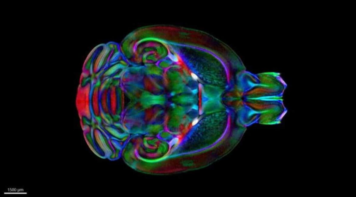
The researchers used diffusion tensor imaging and MRI to capture three-dimensional images of five postmortem mouse brains at the highest resolution ever measured ( 15 microns ), according to Johnson.
The Duke Center for In Vivo Microscopy’s innovative imaging techniques and equipment have made it possible for the researchers to get these pictures at a quality 2.4 million times higher than diagnostic Tests, which the experts have developed over the past 40 years.  ,  ,
To identify important “boney landmarks,” they combined these images with microCT scans of the mouse skull. Finally, they removed the skull’s brains to make it possible to map cells in the same space using light sheet microscopy.  ,  ,
One of the most comprehensive maps of the mouse brain ever created is made possible by the combination of all three methods, which are at the highest spatial resolution in the same space.  ,  ,
About this news from brain mapping research
Author: Shantell Kirkendoll
Source: Duke University
Contact: Shantell Kirkendoll – Duke University
Image: The image is credited to Duke University
Open access to original research.
G. Allan Johnson and colleagues ‘” The Duke Mouse Brain Atlas: MRI and Light Sheet Microscopy Stereotaxic Atlas of the Mouse Brain” Science Advances
Abstract
The mouse brain atlas is a stereotaxic atlas created by MRI and light sheet microscopy.
Atlases of the brain are essential tools for sharing information in a common reference frame.
Unexpectedly, there is no three-dimensional ( 3D ) stereotaxic atlas of the mouse brain that covers the entire brain from macro to single-cell to three-cell levels.
The highest resolution ever recorded for a perfusion-fixed ( in skull ) specimen was 15 micrometers of diffusion tensor images, which were taken at the highest resolution.
Diffusion tensor imaging produces several 3D volumes, each of which highlights distinctive cytoarchitecture.
To create external landmarks ( bregma and lambda ), the averages were mapped into micro-computted tomography of the mouse skull. Coregistered light sheet images of the same brains provided cell maps in the same stereotaxic space.
The Allen Reference Atlas was registered as the volume to correct the geometrical erasure and bring it into stereotaxic space.
The 13 terabytes-long multiscalar atlas provides a common spatial framework for annealing data from molecular, structural, and functional studies of mice.
