Summary: A recently developed PET radiotracer, 18F-PDE-1905, targets a crucial protein within the body’s immune cells to provide high-resolution scanning of neuroinflammation. This book sensor concentrates on PDE4B, a crucial regulation of inflammation signaling, in contrast to standard tracers that target standard downstream markers.
The sensor produced clearer pictures and a wider head distribution than the current TSPO-targeting reflectors in mouse versions of Parkinson’s and multiple sclerosis. This development might lead to earlier, more precise treatment and help to develop precision-guided treatments for neurological conditions like Alzheimer’s, Und, and MS.
Important Information
- PDE4B, a crucial protein in microglial swelling, is instantly targeted by 18F-PDE-1905.
- In terms of quality and coverage, the tracer outperformed regular TSPO tracers.
- Clinical Potential: Promotes personal care, treatment planning, and early detection of neurological diseases.
Origin: SNMMI
According to studies presented at the Society of Nuclear Medicine and Molecular Imaging 2025 Annual Meeting, a recently developed Canine radiotracer has demonstrated the ability to produce high-quality pictures of real-time brain disease.
The sensor may help to advance the detection and more accurate care of a range of neurological conditions, including Alzheimer’s, Parkinson’s, ALS, and multiple sclerosis by identifying activated defense cells in the brain.
Neuroinflammation, an immune reaction brought on by infection, toxin accumulation, or injury to the central nervous system, is a major factor in the development of many neurological and psychiatric disorders. Neuroinflammation scanning plays a crucial role in treatment, monitoring, and treatment.
According to Jiahui Chen, PhD, associate professor in the Department of Radiology and Imaging , Sciences , Emory University School of Medicine in Atlanta, Georgia,” current clinical Dog scanning for neuroinflammation primarily uses tracers that specific TSPO, a river symbol that is broadly expressed across multiple mobile types.”
” Our study presents 18F-PDE-1905, a novel PET tracer that was created specifically to target phosphodiesterase 4B ( PDE4B), a crucial intracellular enzyme that controls inflammatory signaling within microglia, the immune cells of the central nervous system.”
To assess PDE4B expression in neuroinflammatory diseases, researchers began by using bioinformatics tools to analyze a genomics database.
The 18F-PDE-1905 was used alongside the TSPO-specific tracer 18F-D2-LW223, who developed a mouse model of neuroinflammation and performed dynamic PET imaging.
To evaluate protein expression and confirm its association with the PET imaging findings, follow-up analyses were carried out.
PDE4B levels were found to be higher in both Parkinson’s disease and multiple sclerosis patients as well as in corresponding mouse models.
In contrast to controls, PET imaging revealed a significantly higher uptake of 18F-PDE-1905 in diseased mice, indicating that more tracer activity is occurring in neuroinflammatory conditions.
18F-PDE-1905 demonstrated superior image quality and greater brain distribution, which supports its promise for imaging neuroinflammation, in contrast to the TSPO-specific tracer 18F-D2-LW223.
18F-PDE-1905, which targets PDE4B directly, provides a more precise and upstream view of microglial activation, which Chen said is an important early and crucial component of the progression of many neurological diseases.
This could lead to earlier and more precise diagnoses, better monitoring of treatment effectiveness, and more personalized therapies based on direct neuroinflammation measurements, according to the patient.
In the end, 18F-PDE-1905 has the potential to lead to a significant shift toward precision-guided care in neurodegenerative disorders.
About this research in neuroimaging and inflammation
Author: Rebecca Maxey
Source: SNMMI
Contact: Rebecca Maxey – SNMMI
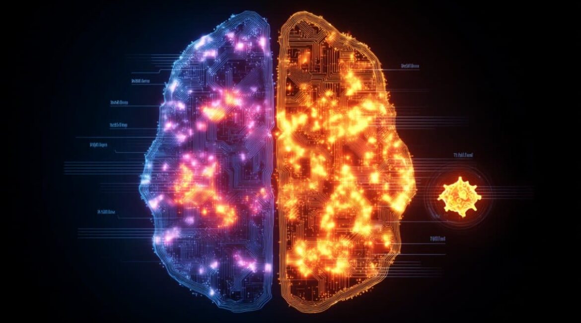
Image: The image is credited to Neuroscience News
The results of original research will be presented at the 2025 Annual Meeting of the Society of Nuclear Medicine and Molecular Imaging.
