Summary: A book PET scanning study has discovered fascinating neuroinflammation patterns in patients with liberal apraxia of discourse ( PAOS), a rare disorder that impairs the body’s capacity to organize conversation. Researchers investigated the presence of increased swelling in the action and speech regions of the brain, especially in those with Parkinson-plus illnesses, using TSPO PET scans.
The research links tau disease and illness severity in the brain, suggesting that inflammation might be a key factor in PAOS progression. This information opens up new avenues for tau-related neurological diseases analysis, tracking, and therapeutic targeting.
Important Information:
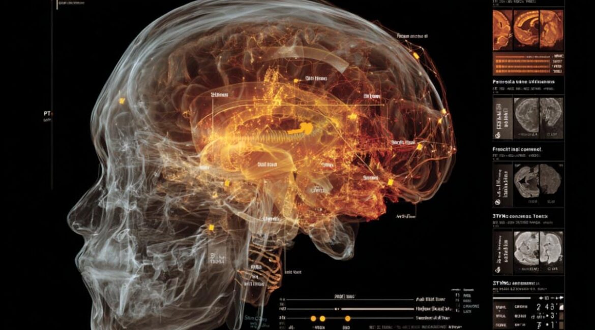
- Disease Hotspots: PET scans revealed swelling in the premotor cortex, frontal lobes, and midbrain, as well as in motor and speech areas.
- Parkinson-Plus Link: Patients with Parkinson-plus characteristics had a greater level of infection and a stronger association with tau deposition.
- Possible diagnostic: Neuroinflammation may aid in the development of PAOS and inform future treatment options.
Origin: SNMMI
In people with liberal apraxia of speech ( PAOS), a rare neurodegenerative disorder that affects speech planning, distinct patterns of brain disease have been identified using a book PET scanning technique.
These results provide new information into how beta disease and neuroinflammation may contribute to PAOS disease progression, opening up new avenues for earlier diagnosis and treatment.
This study was presented at the 2025 Annual Meeting of the Society of Nuclear Medicine and Molecular Imaging.
PAOS is a neurological condition that impairs the ability of the brain to organize and organize speech. It is characterized by a gradual speaking rate, twisted sounds, and laboriously repetitive facial movements when speaking.
Patients with PAOS are more likely to have Parkinson-plus symptoms in the early stages, meet the requirements for democratic supranuclear paralysis or corticobasal symptoms, and usually have a 4-repeat tauopathy at autopsy.
The geographical patterns of neuroinflammation, and how they relate to Parkinson-plus syndromes and tau accumulation, are still terribly understood, according to Ryota Satoh, associate professor at the Mayo Clinic in Rochester, Minnesota, despite earlier neuroscience studies showing significant brain hypertrophy and tau accumulation in the premotor cortex and subcortical regions of PAOS patients.
” Our study aimed to chart the spatial styles of neuroinflammation in PAOS, both with and without Parkinson-plus features, and to investigate how infection interacts with tau testimony.”
In order to measure brain inflammation and tau buildup, the study included 25 PAOS patients ( 13 of whom had Parkinson-plus syndrome ) and 30 healthy controls.
A uniform head atlas was used as a reference point for the study’s analysis of 84 brain regions to determine how swelling and tau PET signals were related to each patient. Then they compared these ranges between patients and good controls and looked at how inflammation and tau were related to various brain regions.
Patients with PAOS were found to have more mental disease than good controls, especially in the areas of the brain that control motion and speech, such as the premotor brain, frontal lobes, basal ganglia, and midbrain, according to 11C-ER176 TSPO Dog scans.
Patients with Parkinson-plus syndrome had wider uptake patterns and higher correlations than those without the syndrome, which suggests that severe neuroinflammation is linked to the presence of Parkinson-plus syndrome.
These findings may help us better understand the neuroinflammatory process that drives PAOS and give us potential applications as a disease biomarker, Satoh said. We think our findings may have a positive impact on the development of neuroinflammatory PET techniques.
About this news from neuroimaging research
Author: Rebecca Maxey
Source: SNMMI
Contact: Rebecca Maxey – SNMMI
Image: The image is credited to Neuroscience News
The results of original research will be presented at the 2025 Annual Meeting of the Society of Nuclear Medicine and Molecular Imaging.
