Summary: New research shows that low-intensity repetitive transcranial magnetic stimulation (rTMS ) can restore key synaptic structures in mouse models of Alzheimer’s disease. The study found that axonal boutons—sites where neurons form connections—had reduced churn in Alzheimer’s animals, indicating affected brain plasticity.
After a second rTMS period, the attrition of one signals type considerably increased, matching levels seen in good mice. These changes suggest that rTMS may partly reverse neural deficits, possibly improving mental connectivity.
Important Information:
- Neural Turnover Boost: rTMS increased churn of terminaux boutons by away to 213 % in AD animals.
- Careful Answer: Just terminaux boutons, no en translates boutons, responded to rTMS.
- Medical Potential: rTMS restored impaired neural flexibility to near-healthy levels.
Origin: SPIE
Alzheimer’s disease ( AD ) is a debilitating neurodegenerative condition that affects a significant proportion of older people worldwide.
Connections are points of contact between neuronal cells that are pliable to change based on our activities. By adding, removing, strengthening, or weakening neural connections, our mental encodes new events or remembers earlier ones.
In AD, neural flexibility, the brain’s ability to regulate the strength of neural connections between neurons, is tremendously disrupted. This increases over time, reducing mental and memory capabilities leading to reduced quality of life. To date, there is no effective solution for Campaign, and only minimal solutions for managing the symptoms.
Studies have shown that repetitive transcranial magnetic stimulation (rTMS ), a noninvasive mental excitement technique that uses electric pulses to target specific brain regions, has medicinal ability to control dementia and associated diseases.
From previous research, we know that rTMS may promote neural plasticity in good nervous systems. Also, it is already used to handle certain neurological and neurological conditions. But, individual reactions to rTMS for Advertising management are changing, and the actual mechanisms are not clearly understood.
Recently, researchers from the University of Queensland ( Australia ) and the Wicking Dementia Research and Education Centre at the University of Tasmania investigated the effects of rTMS on synapses in the brain cortex of mice with Alzheimer’s type dementia.
Their report is published in Neurophotonics. “Since synaptic dysfunction is a key mechanism in AD, in this study, we quantified the changes in synaptic axonal boutons in AD mouse model in response to rTMS, comparing them to those in healthy mice, ” explains corresponding author Dr. Barbora Fulopova, a professor at University of Queensland.
Axonal boutons are specialized endings of an axon, which is the long slender part of a neuron that connects neurons by transmitting neural signals. These are sites where synapses form, allowing neurons to communicate.
Therefore, any change in the number or function of these boutons can have profound effects on brain connectivity. In this study, the researchers observed structural changes of two types of excitatory boutons , namely “terminaux boutons ” ( TBs ) ( short protrusions from the axon shaft typically connecting neurons in a local area ) and “en passant boutons ” ( EPBs ) ( small bead-like structures along axons typically connecting distal regions ). They used two-photon imaging to visualize individual axons and synapses in the brain of a live animal.
The study was conducted on the APP/PS1 xThy-1GFP-M strain of mice, which is a cross between the APP/PS1 strain ( genetically modified to show AD-like symptoms seen in humans ) and the Thy1-GFP-M strain, which expresses a fluorescent protein in certain neurons. This combination causes axons to glow during imaging, enabling precise tracking of synaptic bouton changes over time.
The team monitored the dynamics of the axonal boutons in these mice at 48-hour intervals for eight days, both before and after a single rTMS session. They then compared these findings to healthy wild-type ( WT ) mice.
They found that both TBs and EPBs in the AD mouse model had comparable density to those in healthy WT mice. However, the turnover of both bouton types was significantly lower in the AD mouse model before rTMS, likely due to the amyloid plaque buildup, a key marker of dementia, and potentially causing diseases like AD.
After a single session of low-intensity rTMS, the turnover of TBs in both strains increased significantly, while there was no change in the EPB turnover. Notably, the largest changes were observed two days after stimulation with an 88 percent increase in TB turnover for the WT strain and a 213 percent increase in the APP-GFP strain. However, this increase returned to pre-stimulation levels by the eighth day.
Furthermore, in the AD mouse model, this increased turnover was comparable to the turnover levels in the WT mice seen before stimulation. This indicates that low-intensity rTMS can potentially restore the synaptic plasticity of TBs to those seen in healthy mice.
Moreover, the fact that only TBs, and not EPBs, responded to rTMS points to the possibility that the mechanisms of rTMS might be cell-type specific.
“This is the first study to provide evidence of pre-synaptic boutons responding to rTMS in a healthy nervous system as well as a nervous system marked by the presence of dementia, ” remarks Fulopova.
“Given the established link between synaptic dysfunction and cognitive decline in dementia and the use of rTMS for the treatment of other neurodegenerative conditions, our findings highlight its potential as a powerful addition to currently used AD management strategies. ”
This study marks a significant step forward in understanding AD. While further research is required, the findings of this study pave the way for targeted rTMS treatments that could improve the quality of life of patients with Alzheimer’s disease.
About this brain stimulation and Alzheimer’s disease research news
Author: Daneet Steffens
Source: SPIE
Contact: Daneet Steffens – SPIE
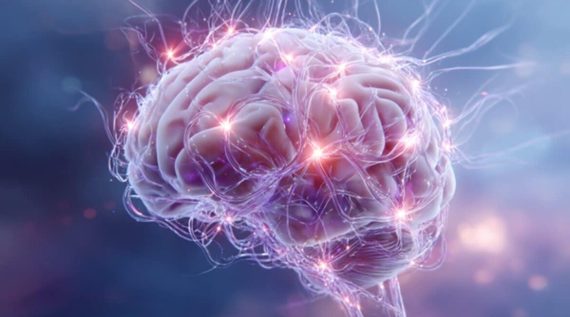
Image: The image is credited to Neuroscience News
Original Research: Open access.
“Repetitive transcranial magnetic stimulation increases synaptic plasticity of cortical axons in the APP/PS1 amyloidosis mouse model ” by Barbora Fulopova et al. Neurophotonics
Abstract
Repetitive transcranial magnetic stimulation increases synaptic plasticity of cortical axons in the APP/PS1 amyloidosis mouse model
Significance
Growing evidence highlights the therapeutic potential of repetitive transcranial magnetic stimulation (rTMS ) in diseases causing dementias such as Alzheimer’s disease ( AD ). However, individual responses to rTMS are variable, and its underlying neural mechanisms are not fully understood.
Aim
As synaptic dysfunction is one of the key mechanisms associated with cognitive deficits in dementia, we investigated the effect of rTMS on cortical synapses using an APP/PS1 amyloidosis mouse model of AD crossed with fluorescent reporters linked to the Thy-1 promoter.
Approach
Using in vivo two-photon imaging, we characterized the plasticity of excitatory terminaux ( TB ) and en passant ( EPB) axonal boutons at 48-h intervals for 8 days on either side of a single session of rTMS.
Results
We found both types of axonal boutons preserved the overall number of their synaptic outputs in wild type ( WT ) and APP/PS1 groups, pre- and post-stimulation. Both synapse types also showed a significantly reduced dynamic fraction in APP/PS1 compared with WT axons pre-stimulation. Following stimulation, the TB, but not EPB, dynamic fraction increased in both WT and APP/PS1 groups.
Conclusions
This suggests possible mechanisms of rTMS action that are cell type-specific and, together with previous findings of improved functional performance, present a potential clinical avenue for rTMS in the management of AD.
