Summary: Researchers have created a 3D-printed implant that promotes muscle regeneration and delivers targeted electric stimulation to cervical cord-damaged areas. In laboratory tests, the implant stimulates neuron and stem cell growth by mimicking the structure of the spinal cord with a fine, electric conductive mesh.
The team improved the effectiveness of the implant by changing the fiber format, opening the door to more potential for medical use. Thanks to the collaboration between technicians, clinicians, and patients, this novel approach could improve treatment for cervical injuries and beyond.
Important Information:
- By delivering electronic signs, a 3D-printed implant encourages nerve repair.
- Neuron growth is boosted by sensitive nanomaterials and personalized fiber patterns.
- Technology might also be applied to cerebral, musculoskeletal, and cardiac healing.
Origin: RCSI
A 3-D published transplant that is used to supply electric stimulation to damaged areas of the spinal cord has been developed by a research group at RCSI University of Medicine and Health Sciences, which could provide a potential new method of recovering brain injury.  ,
Information about the 3-D published transplant and how it performs in laboratory tests have been published in the journal Advanced Science.  ,
Spinal cord injury is a life-changing problem that can cause paralysis, sensory loss, and severe pain. More than 2,300 people and families in Ireland are living with spinal cord injuries, but there is no effective way to treat them.
However, the healing effects of electrical stimulation at the site of the injury have been demonstrated to be potent in promoting the growth of nerve cells ( neurons ).  ,
Our team is developing electric conductive materials that could help the body repair the damaged tissue, according to Professor FergalO’Brien, Deputy Vice Chancellor for Research and Innovation, Professor of Bioengineering and Regenerative Medicine at RCSI, and Head of RCSI’s Tissue Engineering Research Group ( TERG ).” Historically, it has been challenging to promote the regeneration of cells after spinal cord injury.
” The unique surroundings provided by the AMBER Centre provides a significant opportunity for destructive innovation such as this,” said Dr. Martin Luther King, MD.  ,  ,
Researchers from RCSI’s TERG and the Research Ireland Centre for Advanced Materials and Bioengineering Research ( AMBER ) led the study.
The group combined ultra-thin nanomaterials from Professor Valeria Nicolosi’s lab at Trinity College Dublin with 3-D printing to create a smooth gel-like construction using ultra-thin nanomaterials from Professor Valeria Nicolosi’s experiment in the School of Chemistry and AMBER at Trinity College Dublin.  ,
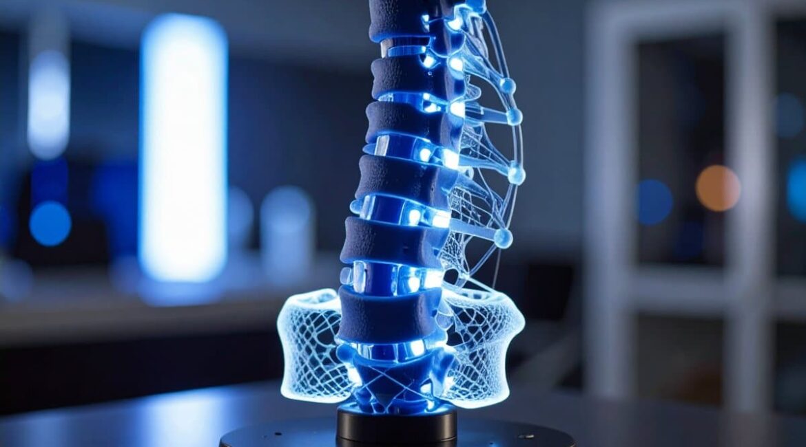
The resulting transplant resembles the structure of the human spinal cord and has a good grid of tiny fibers that can supply power to our tissues. The implant was demonstrated to be capable of effectively delivering electric signals to cells and stem cell, enhancing their capacity to grow when tested in the facility.  ,  ,  ,
Additionally, it was discovered that changing the fiber format of the implant would further increase its effectiveness.  ,
Dr. Ian Woods, Research Fellow at TERG and the study’s second writer, commented,” These 3D-printed components allow us to tune the distribution of electrical stimulation to command renewal.
This technology has the potential to be used in cardiac, orthopaedic, and neurological treatments where electrical signaling can facilitate healing, beyond spinal repair.
The Irish Rugby Football Union Charitable Trust ( IRFU-CT) and the RCSI and AMBER researchers collaborated on the project and created an advisory panel to oversee and guide the research. Rugby players with serious injuries, clinicians, neuroscientists, and researchers made up the group.  ,
The advisory panel “facilitated the development of our understanding of the lived experiences of individuals with spinal cord injuries, their treatment priorities, and emerging treatment approaches,” said Dr. Woods.
Our regular meetings resulted in a constant exchange of ideas, thoughts, and outcomes.
Funding: The Irish Rugby Football Union Charitable Trust, AMBER, the Research Ireland Centre for Advanced Materials and BioEngineering Research, and an Irish Research Council Postdoctoral Fellowship were all partners in funding the study.  ,  ,
About this news about neurotech and SCI research
Author: Laura Anderson
Source: RCSI
Contact: Laura Anderson – RCSI
Image: The image is credited to Neuroscience News
Original research: Free of charge.
The paper “3D printing of electroconductive MXene-based micromeshes in a biomimetic hybridized hyaluronic acid-based scaffold directs and enhances electrical stimulation for neural repair applications” by Ian Woods and colleagues. Advanced Science
Abstract
Electroconductive MXene-based micromeshes in a biomimetic hybridized hyaluronic acid-based scaffold are 3D printed to enhance and direct electrical stimulation for neural repair applications.
Although recent advances in electrical stimulation suggest some promise in neural tissue repair, no effective treatments are currently available for central nervous system neurotrauma.
It is suggested that the structured integration of an electroconductive biomaterial into a scaffold for tissue engineering can enhance electroactive signaling for neural regeneration.
The MAX-phase powder used to create electroconductive 2D Ti3C2Tx nanosheets and MXene nanosheets exhibits excellent biocompatibility with neurons, astrocytes, and microglia.
Melting-electrowriting is used to 3D-print highly-organized PCL micro-meshes with varying fiber spacings ( low, medium, and high-density ), which are functionalized with MXenes to provide highly-tunable electroconductive properties ( 0. 081 0.053-18. 87 2. 94 S/m ) to achieve a spatially-controlled distribution of these MXenes.
A soft, growth-supporting MXene-ECM composite scaffold was created by embedding these electroconductive micromeshes within a neurotrophic, immunomodulatory hyaluronic acid-based extracellular matrix (ECM).
Neurite outgrowth was facilitated by electrical stimulation of neurons seeded on these scaffolds, which was influenced by fiber spacing in the micro-mesh.
In a multicellular model of cell behavior, neurospheres stimulated for 7 days on high-density MXene-ECM scaffolds showed significantly more axonal extension and neuronal differentiation than low-density scaffolds and MXene-free controls.
The findings demonstrate that the spatial organization of electroconductive materials in neurotrophic scaffolds can enhance electrical stimulation repair-critical responses, and that these biomimetic MXene-ECM scaffolds represent a promising new method of neurotrauma repair.
