
Summary: Researchers have developed an innovative system that can detect glioblastoma, an extreme brain tumor, in under an hour using a book biochip. From a small blood sample, the chip uses electrokinetic technology to identify active Epidermal Growth Factor Receptors ( EGFRs ) in extracellular vesicles.
This approach has great sensitivity and selectivity, reducing interference and possibly enhancing early detection. The technology could be used to improve the effectiveness of its clinical applications by identifying additional conditions.
Important Information:
- New system symptoms glioblastoma in less than 60 days.
- uses an electrokinetic biochip to identify blood samples that contain engaged EGFRs.
- Prospective for adaptation to treat other conditions besides glioblastoma.
Origin: University of Notre Dame
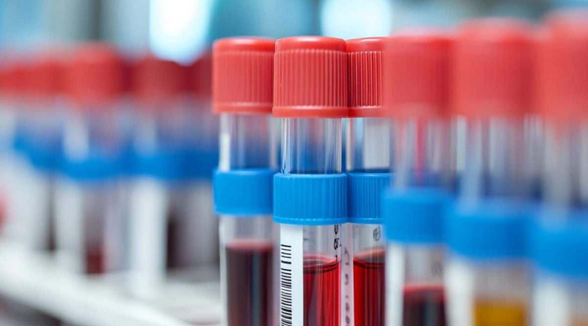
Researchers at the University of Notre Dame have developed a book, automated machine capable of diagnosing glioblastoma, a fast-growing and irreversible brain tumor, in less than an afternoon. The typical glioblastoma individual survives 12-18 months after treatment.
A biochip that uses electrokinetic technology to identify biomarkers, or active Epidermal Growth Factor Receptors ( EGFRs ), that are overexpressed in some cancers, such as glioblastoma, and found in extracellular vesicles, is the diagnostic’s center.
Cells or extracellular vesicles are distinctive nanoparticles that cells secrete. They are great — 10 to 50 days bigger than a protein — and they have a weak demand. Our systems was especially designed for these particles, using their features to our benefits”, said , Hsueh-Chia Chang, the Bayer Professor of Chemical and Biomolecular Engineering at Notre Dame and lead author of the , research about the diagnostic , published in , Communications Biology.
The researchers had two goals: creating a medical technology that could detect effective EGFRs on extracellular vesicles while developing a process that could distinguish between active and passive EGFRs.
Experts created a biochip that uses an affordable, electrokinetic device about the size of a game in a ball pen to accomplish this. Antibody on the sensor may form various bonds to the same extracellular vesicle due to the size of the external vesicles. This approach drastically improves the diagnostic’s awareness and sensitivity.
Therefore, while bringing a large negative charge, chemical silica nanoparticles “report” the presence of effective EGFRs on the captured extrinsic vesicles. A voltage change can be observed when there are extracellular vesicles carrying energetic EGFRs, indicating that the patient has glioblastoma.
This charge-sensing technique uses electrochemical reactions or fluorescence to minimize interference that is prevalent in recent sensor technologies.
Our electrokinetic device enables us to perform tasks that conventional diagnostics cannot, according to Notre Dame’s research affiliate professor of chemical and biomedical engineering and co-author of the study.
Because our detector is unaffected by other particles or molecules, we can directly load body without having to wait for a while to remove external vesicles. It shows small noise, making ours more able to detect diseases without the use of other technologies.
In total, the system includes three parts: an technology program, a mockup of a portable system that administers materials to manage the check and the biochip. The technology layout and design are reuseable despite the fact that each test calls for a new biochip.
Running one evaluation takes under an hour, requiring just 100 microliters of body. Each biochip costs less than$ 2 in materials to manufacture.
Although this medical tool was created for glioblastoma, the experts claim that it can be used for other types of natural nanoparticles. This opens up the possibility of the tech detecting a number of different indicators for various illnesses.
Chang stated that the team is looking into the potential for diagnosing ovarian cancer and other conditions like diabetes, cardiovascular disease, dementia, and epilepsy.
Because of how dangerous it is and the absence of early screening tests, Chang said,” Our strategy is no specific to glioblastoma, but it was specifically appropriate to begin with it.” ” Our desire is that if early recognition is more appropriate, then there is an improved chance of survival”.
The Olivia Newton-John Cancer Research Institute’s Centre for Research on Brain Cancer provided blood tests for the device’s assessment, which was conducted in Melbourne, Australia.
Other researchers include Andrew Scott and Hui Gan from the Olivia Newton-John Cancer Research Institute and La Trobe University, as well as previous complete at Notre Dame Nalin Maniya and Sonu Kumar, Jeffrey Franklin, James Higginbotham, and Robert Coffey from Vanderbilt University.
Funding:
The National Institutes of Health Common Fund provided funding for the review.
About this information on brain tumor study
Author: Brandi Wampler
Source: University of Notre Dame
Contact: Brandi Wampler – University of Notre Dame
Image: The image is credited to Neuroscience News
Original Research: Start entry.
Hsueh-Chia Chang et al.,” An electrolyte trade barrier sensor detects EGFR and its task state in blood CD63 external vesicles from people with glioblastoma.” Communications Biology
Abstract
In plasma CD63 extracellular vesicles from patients with glioblastoma, an anion exchange membrane sensor detects EGFR and its activity state.
We present a quantitative sandwich immunoassay for CD63 Extracellular Vesicles ( EVs ) and a constituent surface cargo, EGFR and its activity state, that provides a sensitive, selective, fluorophore-free and rapid alternative to current EV-based diagnostic methods.
A hydrophilic anion exchange membrane that is detected by detection antibodies and a charged silica nanoparticle reporter that is detected by detection antibodies are used in our sensing design.
This hydrophilic design with charged reporters minimizes interference from dispersed proteins, enabling direct plasma analysis without the need for EV isolation or sensor blocking, while increasing sensitivity and robustness by the ion-depletion action of the membrane.
With a LOD of 30 EVs/μL and a high relative sensitivity of 0.01 % for targeted proteomic subfractions, our assay enables accurate quantification of the EV marker, CD63, with colocalized EGFR by an operator/sample insensitive universal normalized calibration. To demonstrate this new platform, we analyzed untreated clinical samples of glioblastoma.
Notably, we target both total and “active” EGFR on EVs, with a monoclonal antibody mAb806 that recognizes a normally hidden epitope on overexpressed or mutant variant III EGFR.
Analysis of samples yielded an area-under-the-curve ( AUC) value of 0.99 and a low p-value of 0.000033, surpassing the performance of existing assays and markers.
