
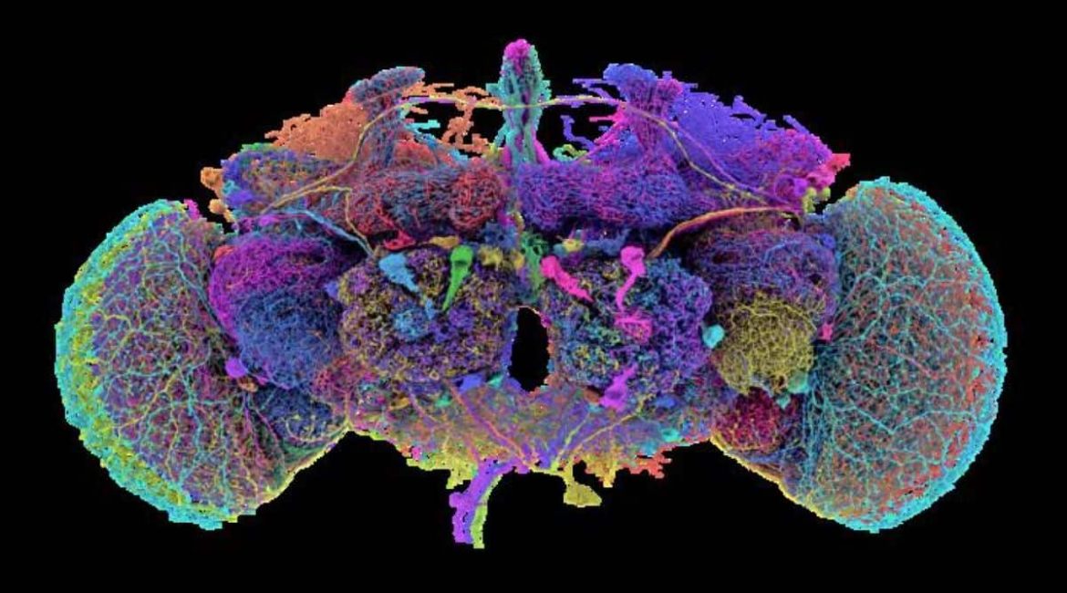
Summary: Researchers have created the first neuron-by-neuron image of an older fruit fly head, marking a major achievement in mental mapping. The image, built using 21 million images, connects almost 140, 000 neurons and 50 million synapses, making it the most complex mental image of any child creature to date.
This” connectome” provides a roadmap for understanding neural circuits and behaviors in more complex species, including humans. The creation, powered by AI, opens doors for fresh insights into mental function and conditions.
Important Information:
- The mature fruit fly mind contains 140, 000 cells and 50 million connections.
- Using AI to practice neurological information, 21 million pictures were used to create the image.
- Fruit flies share 60 % of people Genome, aiding research on human brain disorders.
Origin: Princeton
A Princeton-led team of scientists has built the first neuron-by-neuron and synapse-by-synapse roadmap through the brain of an adult fruit fly ( Drosophila melanogaster ), marking a major milestone in the study of brains.
This research is the premier content in the Oct. 2 particular matter of , Nature, which is devoted to the fresh fruit travel” connectome”.
Earlier researchers mapped the head of a , C. elegans , worm, with its 302 neurons, and the brain of a juvenile fruit fly, which had 3, 000 neurons, but the adult fruit fly is various orders of magnitude more difficult, with about 140, 000 neurons and almost 50 million synapses connecting them.  ,
Fruit flies make up 60 % of human DNA, and three in four of the human genetic diseases are caused by fruit flies. Understanding the brains of larger, more complex species, like humans, is a steppingstone to understanding theirs.
” This is a major achievement”, said Mala Murthy, director of the Princeton Neuroscience Institute and, with , Sebastian Seung, co-leader of the research team. There is no other adult animal with this complexity with a full brain connectome. Murthy is also Princeton’s Karol and Marnie Marcin ‘ 96 Professor of Neuroscience.
Princeton’s Seung and Murthy are co-senior authors on the flagship paper of the , Nature , issue, which includes a suite of nine related papers with overlapping sets of authors, led by researchers from Princeton University, the University of Vermont, the University of Cambridge, the University of California-Berkeley, UC-Santa Barbara, Freie Universität-Berlin, and the Max Planck Florida Institute for Neuroscience.
The work was funded in part by the NIH’s , BRAIN Initiative, the Princeton Neuroscience Institute’s Bezos Center for Neural Circuit Dynamics and McDonnell Center for Systems Neuroscience, and other public and private neuroscience institutes and funds, listed at the end of this document.  ,
The FlyWire Consortium, which is based at Princeton University and consists of teams working in more than 76 laboratories and hosting 287 international researchers as well as volunteer gamers, created the map.  ,
Sven Dorkenwald, the lead author on the flagship Nature paper, spearheaded the FlyWire Consortium.
” What we built is, in many ways, an atlas”, said Dorkenwald, a 2023 Ph. D. graduate of Princeton and currently employed by the University of Washington and the Allen Institute for Brain Science.
You do n’t want to explore your brain without a map, just like you would n’t want to drive to a new location without Google Maps. What we have done is build an atlas of the brain, and added annotations for all the businesses, the buildings, the street names. Researchers now have the tools to thoughtfully navigate the brain as we try to understand it as a result of this.
The fly connectome demonstrates connections between the fruit fly brain at every scale, just like a map that traces out every tiny alley and superhighway.
The map was built from , 21 million images , taken of a female fruit fly brain by a team of scientists led by Davi Bock, then at the Howard Hughes Medical Institute’s Janelia Research Campus and now at the University of Vermont.
The lumps and blobs in those images were transformed into a three-dimensional labeled map using an AI model created by Princeton’s Sebastian Seung and other researchers and software engineers. The researchers immediately made their in-progress neural map available to the scientific community in order to protect their data from being kept secret.  ,
” Always making advances in AI computing have made it possible to map the entire brain. It would not have been possible to manually reconstruct the entire wiring diagram. This is a display of how AI can move neuroscience forward,’ said Prof.  , Sebastian Seung,  , one of the co-leaders of the research and Princeton’s Evnin Professor in Neuroscience and , a professor of computer science.  ,
” Now that we have this brain map, we can close the loop on which neurons relate to which behaviors,” said Dorkenwald.
The advancement might lead to specially designed treatments for brain disorders.  ,
” In many respects, it ( the brain ) is more powerful than any human-made computer, yet for the most part we still do not understand its underlying logic,” said John Ngai, director of the U. S. National Institutes of Health’s BRAIN Initiative, which provided partial funding for the FlyWire project.
Without a thorough understanding of how neurons interact with one another, we wo n’t have a basic understanding of what happens in a healthy brain or what malfunctions in disease.
Funding: This research was supported by the National Institutes of Health ( NIH) BRAIN Initiative ( RF1 MH117815, RF1 MH129268, 1RF1MH120679-01 and U24 NS126935 ) and National Institute Of Neurological Disorders And Stroke ( NINDS ) ( RF1NS121911 ), the Princeton Neuroscience Institute’s Bezos Center for Neural Circuit Dynamics and McDonnell Center for Systems Neuroscience, Google, the Allen Institute for Brain Science, the National Science Foundation ( NSF Neuronex2 2014862, Neuronex2 MRC MC_EX_MR/T046279/1, MRC MC-U105188491, PHY-1734030 ),  , Wellcome Trust Collaborative Award ( 203261/Z/16/Z and 220343/Z/20/Z ), a Marie Skłodowska-Curie postdoctoral fellowship ( H2020-WF-01-2018-867459 ), the Portuguese Research Council ( Grant PTDC/MED-NEU/4001/2021 ), and the Intelligence Advanced Research Projects Activity ( IARPA ) via the Department of Interior ( DOI ) ( D16PC0005 ).
About this news from brain mapping research
Author: Liz Fuller-Wright
Source: Princeton
Contact: Liz Fuller-Wright – Princeton
Image: The image is credited to Tyler Sloan and Amy Sterling / FlyWire / Princeton University
Original Research: Open access.
Mala Murthy and colleagues ‘” An adult brain’s neuronal wiring diagram.” Nature
Abstract
An adult brain’s neuronal wiring diagram
By analyzing and analyzing electron microscopic brain images, it is possible to map connections between neurons. This method has been used to reconstruct local connectivity maps that are highly informative but are insufficient for understanding brain function more broadly in recent years.
Here we present a neuronal wiring diagram of a whole brain containing 5 × 10⁷ , chemical synapses , between 139, 255 neurons reconstructed from an adult female , Drosophila melanogaster. Additionally, the resource includes predictions of neurotransmitter identities, annotations of cell classes and types, nerves, hemilineages, and annotations of cell classes and types.
Data products have been made to work with other fly data resources and are accessible for download, programmatic access, and interactive browsing.
We use the connectome to trace synaptic pathways and the measure of information flow from inputs ( sensory and ascending neurons ) to outputs ( motor, endocrine, and descending neurons ) across both hemispheres and between the central brain and the optic lobes. We also create a projectome, a map of projections between regions, from the connectome.
How structure can uncover putative circuit mechanisms underlying sensorimotor behaviors can be demonstrated by tracing from a subset of photoreceptors to descending motor pathways.
The open ecosystem and technologies described here provide the foundation for larger-scale connectome projects in other species.
