
Summary: To improve the accuracy of 3T MRI photos and provide more information for the detection of brain abnormalities, researchers have created a machine learning model that compares to the higher-resolution 7T MRI images. Finer details, such as light issue lesions and subcortical microbleeds, are often obscured by the manufactured 7T images by conventional MRI systems.
Although clinical validation is required before use, this AI-driven approach may increase diagnostic accuracy for conditions like traumatic brain injuries ( TBI ) and multiple sclerosis ( MS ). Without the use of specific products, the new design may eventually give more people access to high-quality scanning insights. This development makes a convincing connection between medical imaging technology and AI.
Major Information
- 3T MRIs are enhanced by the AI type to closely resemble 7T MRIs in terms of detail.
- Chemical 7T images showed sharper mental disease boundaries, aiding diagnosis.
- By enhancing the representation of mental abnormalities, the model might be beneficial for TBI and MS patients.
Origin: UCSF
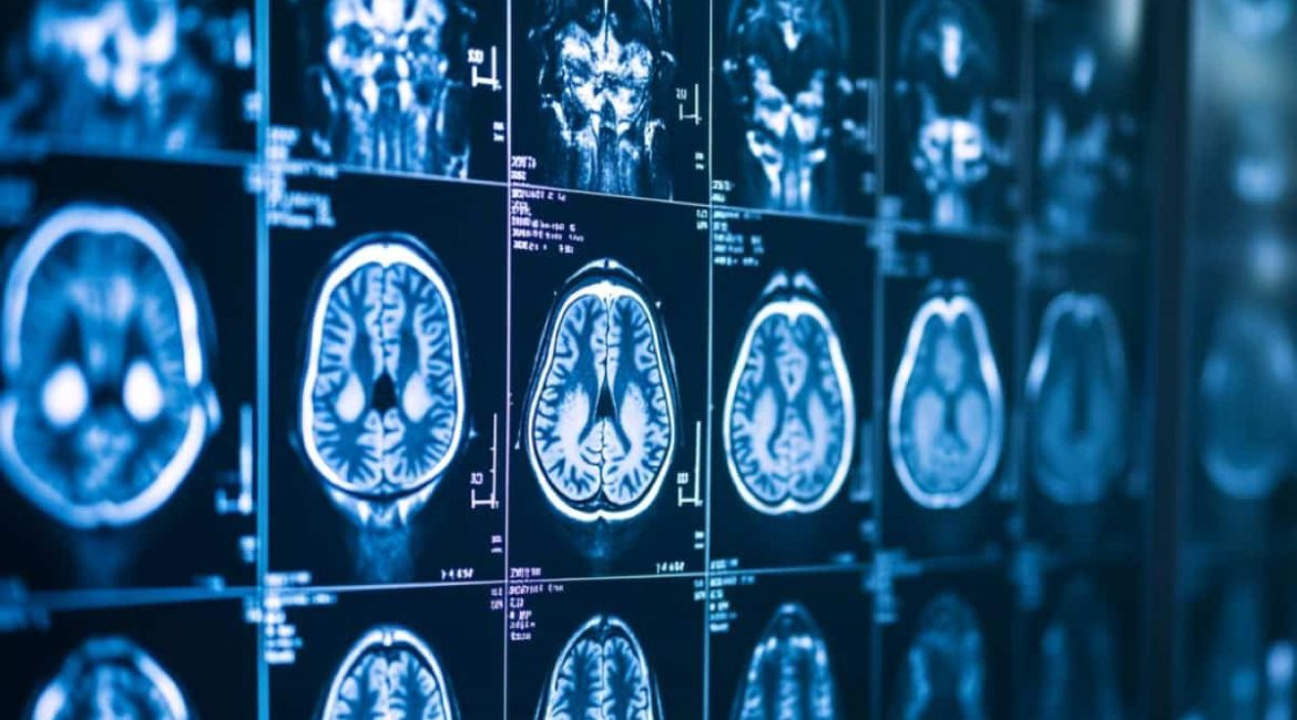
There is growing interest in using machine learning to enhance the imaging data captured by magnetic resonance imaging ( MRI ) technology at the intersection of AI and medicine.
In terms of identifying and monitoring pathological tissue, especially in the brain, ultra-high-field MRI at 7 Tesla (7T ) may have significantly greater resolution and clinical advantages over high-field MRI at 3T in terms of defining anatomical structures.
Most medical MRI examination in the U. S. are performed with 1.5T or 3T MRI techniques. Only about 100 7T MRI models were documented by the National Institutes of Health as of 2022 when they were first documented for use in medical imaging around the world.
By combining 7T-like images with genuine 7T MRIs, researchers at UC San Francisco created a machine learning algorithm to improve 3T MRIs.
Their model, which improved compulsive tissue for better scientific insights, is a first step in the development of synthetic 7T MRI models for clinical applications.
The study was  , presented , Oct. 7 at the , 27th International Conference on Medical Image Computing and Computer Assisted Intervention , ( MICCAI ).
” Our report introduces a machine-learning type to generate high-quality Tests from lower-quality images. We demonstrate how this AI system makes it easier to visualize and recognize brain abnormalities that MRIs intraumatic brain injuries ca n’t do,” said senior study author Reza Abbasi-Asl, Ph.D. D., UCSF Assistant Professor of Neurology.
Our results demonstrate the potential of AI and machine learning to enhance the quality of health photographs captured by less sophisticated imaging systems.
Better to compare various disease and TBI with one another
UCSF researchers collected imaging data from patients diagnosed with mild , traumatic brain injury , ( TBI ) at UCSF. The generated synthetic-7T MRIs, which were created from normal 3T MRIs, were created using three neural network models that had been created to conduct image improvement and 3D image segmentation.
Patients with mild TBI received increased pathological cells from the images created with the new models. They chose to use as an instance an example area with white problem lesions and microbleeds in subcortical regions as a assessment.
Compulsive muscle was apparent in 7T images that had been created more easily. This was clearly seen in the sharper outlines of subcortical microbleeds and the separation of adjacent tumors.
Also, the manufactured 7T pictures better captured the varied features within whitened matter lesions. These studies also highlight the potential for using this technology to increase the diagnostic accuracy in several sclerosis-related neurological disorders.
Although machine learning framework synthetization techniques demonstrate outstanding performance, their application in medical settings will require substantial validation.
Future research should include thorough diagnostic evaluation of the model’s findings, medical rating of the model-generated images, and measurement of uncertainties in the design, according to the researchers.
About this information about analysis in neuroimaging and AI
Author: Reza Abbasi-Asl
Source: UCSF
Contact: Reza Abbasi-Asl – UCSF
Image: The image is credited to Neuroscience News
