
Summary: Researchers have discovered a new type of brain body that can produce energy feelings, refuting the long-held notion that cells are the only ones capable of doing this. Both brain tumors and good human brains both harbored these hybrid cells that are both neuronal and glial.
This breakthrough discovery, made possible through innovative single-cell examination techniques, could provide insights into brain development and normal brain function. These electric effective cells may even have an impact on patient outcomes by their presence in tumors.
Important Facts:
- Composite neuron-glia cells found in cancer can fire electrical impulses.
- These cells can be found in both good human brains and tumors.
- Greater numbers of these rising cross gliomas does indicate better success outcomes.
Origin: Baylor College of Medicine
A fresh body form in the human mind has been discovered thanks to researchers at Baylor College of Medicine and Texas Children’s Hospital’s Jan and Dan Duncan Neurological Research Institute.
The study published in , Cancer Cell , reveals that a third of the tissue in glioma, a type of mental tumor, fire electrical impulses. Ironically, the impulses, also called action potentials, originate from lesion cells that are part nerve and component glia, supporting the groundbreaking idea that neurons are not the only cells that can make electronic signals in the brain.
Additionally, the researchers discovered that tissues in the non-tumor people mental exhibit cross neuron-glia traits. The findings emphasize the need to look into the work of these newly discovered cells in both brain and normal mental function.
” Gliomas are the most prevalent tumors in the central nervous system, with an estimated 12, 000 cases diagnosed annually. These lesions have disastrous effects on mental and neurological functions and are universally fatal.
” Past studies have shown that individual survival effects are associated with tumor proliferation and invasiveness, which are influenced by tumor intrinsic and extrinsic elements, including communication between tumor cells and cells that reside in the brain,” said , Dr. Benjamin Deneen , professor and Dr. Russell J. and Marian K. Blattner Chair in the Department of , Neurosurgery, director of the , Center for Cancer Neuroscience, a member of the , Dan L Duncan Comprehensive Cancer Center , at Baylor and a principal analyst at the , Jan and Dan Duncan Neurological Research Institute.
Researchers have previously discovered that neurons communicate with tumors in ways that promote tumor growth and invasiveness and that glioma and nearby healthy neurons communicate with one another.
” We have known for some time now that tumor cells and neurons interact directly,” said first author , Dr. Rachel N. Curry, postdoctoral fellow in pediatrics – neuro oncology at Baylor, who was responsible for conceptualizing the project.
” But one question that stayed in my mind was,” Are cancer cells electrically active?” We needed human samples straight from the operating room to answer this question correctly. This made sure that the cells ‘ biology as they would be present in the brain was as much preserved as possible.
The team used Patch-sequencing, a combination of techniques that integrates whole-cell electrophysiological recordings to measure spiking signals with single-cell RNA-sequencing and analysis of the cellular structure to identify the type of cells, to study the ability of glioma cells to spike electrical signals and identify the cells that produce the signals.
The electrophysiology experiments were conducted by research associate and co-first author , Dr. Qianqian Ma , in the lab of co-corresponding author associate professor of , neuroscience , Dr. Xiaolong Jiang. This cutting-edge method has never been employed before to study human brain tumor cells.
We were” truly surprised” to learn that these tumor cells had unique morphological and electrophysiological characteristics, Ma said. This was something we had never seen before in the mammalian brain.
We carried out all of these analyses on single cells. We assessed each person’s electrophysiological activity. We extracted the content of each cell and sequenced the RNA to find the genes involved in it. This information, according to Deneen, allows us to identify the different types of cells.
We also used dyes to depict each cell’s structural features to color each cell.
The researchers had to create a novel way to analyze this enormous amount of individual data in order to incorporate it.
” To define the spiking cells and determine their identity, we developed a computational tool – Single Cell Rule Association Mining (SCRAM ) – to annotate each cell individually,” said co-corresponding author,  , Dr. Akdes Serin Harmanci,  , assistant professor of neurosurgery at Baylor.
Finding that so many glioma cells are electrically active was a surprise because it contradicted a fundamental theory in neuroscience that says neurons are the only ones that generate electric impulses among all the various types of cells, Curry said.
Some people have suggested that some oligodendrocyte precursor cells ( ODCs ) in a rodent brain may generate electrical impulses, but it has proved challenging to confirm this in humans.
” Our findings demonstrate that other human cells besides neurons can generate electrical impulses.” The electrical contributions of these cells should be further investigated because the adult brain contains an estimated 100 million of these OPCs.
” In addition, the thorough data analyses revealed that the spiking hybrid cells in glioma tumors had characteristics of both neurons and OPC cells,” Harmanci said.
” Interestingly, we found non-tumor cells that are neuron-glia hybrids, suggesting that this hybrid population not only plays a role in glioma growth but also contributes to healthy brain function”.
The findings also point out that the proportion of spiking hybrid cells in glioma may have an impact on the prognostic value, according to co-authors Dr. Ganesh Rao, Marc J. Shapiro Professor and chair of neurosurgery at Baylor.
The data indicates that the better a patient’s survival outcome the better the number of spiking hybrid glioma cells they have. This data is very helpful to patients and their physicians.
” This work is the result of extensive equal collaboration across multiple disciplines – neurosurgery, bioinformatics, neuroscience and cancer modeling – disciplines strongly supported by state-of-the-art groups at Baylor”, Deneen said.
The findings provide a more comprehensive understanding of glioma tumors and normal brain function, as well as a sophisticated bioinformatics pipeline to examine complex cellular populations and potential prognostic implications for patients with this devastating disease.
Other contributors to this work include Malcolm F. McDonald, Yeunjung Ko,  , Snigdha Srivastava, Pey-Shyuan Chin,  , Peihao He, Brittney Lozzi, Prazwal Athukuri, Junzhan Jing, Su Wang, Arif O. Harmanci and Benjamin Arenkiel. The authors are affiliated with one or more of the following institutions: Baylor College of Medicine, the Jan and Dan Duncan Neurological Research Institute at Texas Children’s Hospital and University of Texas Health Science Center, Houston.
Funding: This work was supported by grants from the NIH ( R35-NS132230, R01NS124093, R01CA223388, U01CA281902, R01NS094615, 5T32HL92332-15, F31CA265156, and F99CA274700 ). Further support was provided by NIH Shared Instrument Grants ( S10OD023469, S10OD025240, P30EY002520 ) and CPRIT grant RP200504.
About this news from brain cancer research
Author: Aaron Nieto
Source: Baylor College of Medicine
Contact: Aaron Nieto – Baylor College of Medicine
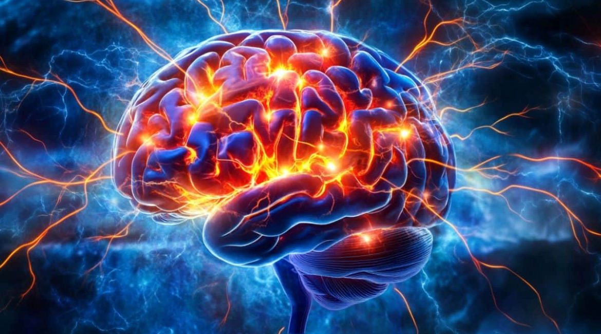
Image: The image is credited to Neuroscience News
Original Research: The findings will appear in Cancer Cell
