
Summary: Researchers have discovered a distinctive mental system that may explain some depressed people. They discovered that people with larger salience systems were more likely to produce depression over time by regularly scanning people ‘ neurons.
This finding suggests that some people may get “pre-wired” for sadness, offering insights into predicting sensitivity to the problem. Although more research is required, this thorough analysis may change the way people with mental illnesses are portrayed and treated.
Important Information:
- The risk of depression may be increased by larger prominence networks in the brain.
- Repeated brain imaging over period revealed this contributing style.
- This method of deep scanning might make it easier to predict psychiatric conditions.
Origin: Weill Cornell University
Researchers at Weill Cornell Medical have discovered a unique style of cerebral interactions that appears to lead some people to despair by regularly scanning the brains of a select group of patients over the course of a year and a half.
Published Sept. 4 in , Nature, the labor highlights the potential of a new “deep scanning” approach to help determine a person’s susceptibility to depression and other neurological conditions and perhaps guide the development of novel treatments.
Functional magnetic resonance imaging ( fMRI ) has been used for decades by neuroscientists to measure changes in blood flow and identify patterns of activity in the brain. This has been a valuable resource for studying adult brain function.
Unique brain activity patterns vary between individuals and over time in a single individual. That’s particularly dangerous for studying problems such as depression.
” Depression is, by definition, an acute psychiatric illness, it’s characterized by periods of low feelings mixed in with intervals of wellness”, says top author , Dr. Conor Liston, professor of psychiatry in the Department of Psychiatry and professor of neuroscience at , Feil Family Brain and Mind Research Institute , at Weill Cornell Medicine.
What are the procedures that govern those changes over time? he asks.
Pre-wired for Unhappiness?
In order to address that, the researchers enrolled a small number of patients who had been diagnosed with depression as well as a larger number of unaffected controls and fMRI scan their brains numerous times over the course of several months.
The salience network, a brain feature that is nearly two-fold larger in most individuals with depression symptoms than in settings who did n’t have clinical depression, was discovered by the deep scanning technique.
A group of frontal lobe and striatum-related head regions are thought to be involved in prize processing and determining which stimuli merit attention.
” Having a larger intensity system appears to increase the risk for depression—the result is an order of magnitude larger than what we often see in ultrasound research”, says Dr. Liston, who is also a physician at NewYork-Presbyterian/Weill Cornell Medical Center.
The scientists extended the study’s scope to include data from hundreds of additional patients whose brains had been scanned less often in collaboration with a sizable group of global collaborators. According to those findings, those who had larger intensity systems in youth were more likely than those who had been programmed for melancholy later in life.
Second Steps
Past research has examined how the body’s processing of rewards is related to the prominence network. Being implicated in despair makes sense, says Dr. Charles Lynch, associate professor of neuroscience in the Department of Psychiatry at Weill Cornell Medicine and lead author of the new study.” One of the main deficits in melancholy is lethargy, which is the inability to feel pleasure and enjoy daily activities,” he says.
The study has already significantly strengthened the deep scanning approach, despite the scientists ‘ claims that the results must be reproduced and extended before they can be used directly in the clinic.
” For years, many investigators assumed that brain networks look the same in everybody”, Dr. Lynch says. However, the findings of this study strengthen existing research that shows that people have fundamental differences.
He adds that the team now hopes to research how various depression medications affect brain networks ‘ activity and might apply their findings to other neuropsychiatric conditions as well.
About this news item about depression and neuroimaging
Author: Krystle Lopez
Source: Weill Cornell University
Contact: Krystle Lopez – Weill Cornell University
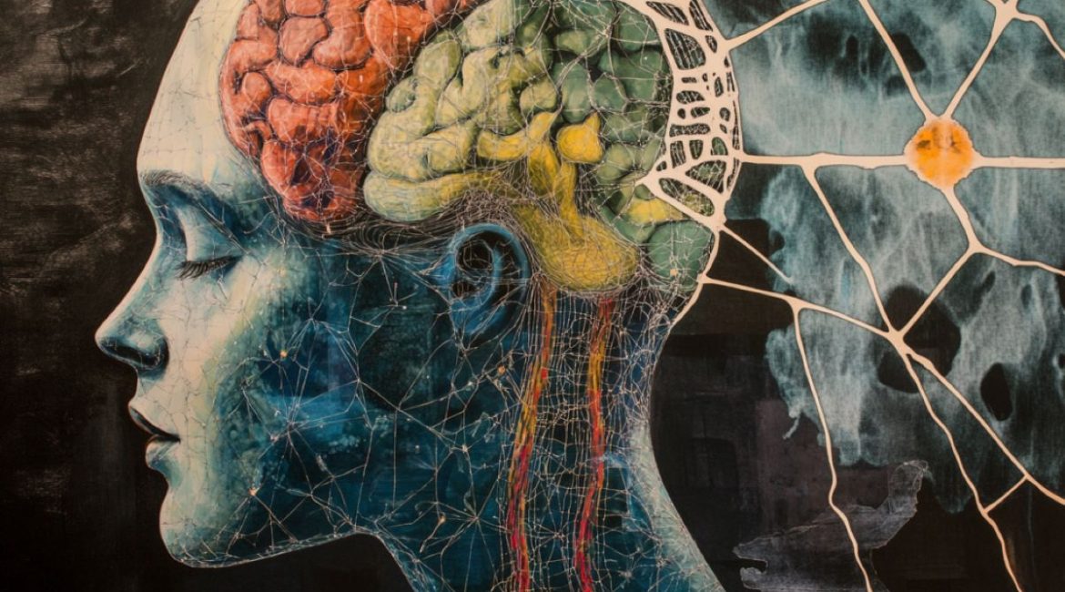
Image: The image is credited to Neuroscience News
Original Research: The findings will appear in Nature
