
Summary: MRI-guided energy, or MRI-linac, you track glioblastoma tumor changes in real-time during radiation treatment. Researchers discovered that daily MRI scans consistently predicted growth in 26 % of cases and accurately matched tumor changes detected by conventional imaging 74 % of the time.
This technology could help for quicker treatment adjustments, improving patient outcomes. The study highlights MRI-linac as a promising treatment for brain tumor by revealing accurate data on tumor conduct in real-time.
Important Information:
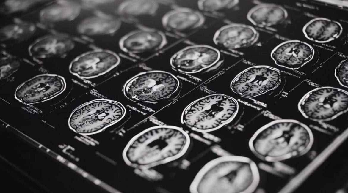
- During energy therapy, glioblastoma lesions are monitored by MRI-linac systems.
- In 74 % of cases, daily MRI scans delivered comparable results to conventional imaging.
- MRI-linac was signal tumor growth before, enabling faster therapy adjustments.
Origin: University of Miami
A recent study demonstrates the potential energy of imaging combined with energy to influence glioblastoma people ‘ care in real time.
The research, led by researchers at , Sylvester Comprehensive Cancer Center, part of the University of Miami Miller School of Medicine,  , is the first to estimate tumor changes in glioblastoma clients receiving MRI-guided radiation treatment.
This book technique, also known as MRI-linear throttle or MRI-linac, sets regularly imaging with radiation. One of the few tumor centers to provide this technology to patients with glioblastoma is Sylvester, which was the first to do so.
The Sylvester researchers discovered that this routine imaging can be used as a crucial monitoring tool to detect tumor growth before starting treatment, as well as conventional imaging, giving proof that the technique might one day be used to aid in speedy treatment adaptation during radiation therapy.
The study was published Sept. 30 in the , International Journal of Radiation Oncology – Biology – Physics , and simultaneously presented at the American Society for Radiation Oncology ( ASTRO ) meeting by the study’s first author, Kaylie Cullison, Ph. D., an M. D. /Ph. D. student in the Miller School ‘s , Medical Scientist Training Program.
According to Cullison,” Our study indicates that these normal scans can provide an early warning sign for possible tumor growth.”
In the study, which was led by , Eric A. Mellon, M. D., Ph. The experts followed 36 glioblastoma people over a six-week program of regular energy and MRI scans using MRI-linac, led by Dr. Sylvester, a radiation oncology and co-leader of Sylvester’s Neurologic Cancer Site Disease Group.
A second MRI picture with distinction taken one week before the radiation treatment and another one a fortnight after the rays course is finished, the data from these everyday scans were then compared to standard-of-care imaging to determine tumor size.
Some patients are concerned about regular use of these heavy metals, but the study was conducted without distinction for the regular scans, even though MRI-linac may be paired with a contrast agent.
To accurately position the patient under the rays beam, the patient is usually guided by an X-ray or CT scan. However, these types of imaging merely show where the bones is located.
” Any appliance that does not include MRI, which is 99 % of radiation-delivery products out there, cannot see what’s going on inside of the mind”, Mellon said. ” MRI-linac allows you to view what’s going on in the head, for the first time”.
The scientists found that for 74 % of the test individuals, their MRI-linac scanning matched the information found in the common, compare MRI performed before and after therapy.
In other words, both approaches agreed on whether the clients ‘ cancers grew, shrank, or remained the same size throughout the course of radiation treatment. For the other 26 % of patients, the MRI-linac imaging predicted tumor growth while the pre- and post-treatment imaging showed that the tumor shrank.
Although the normal MRI did certainly always agree with the comparison imaging, the fact that it did not lose any real lesion progress suggests that it could be used to predict potential tumor development during radiation therapy, the scientists said.
That message could then be confirmed with contrast imaging and, finally, could be used to modify the patient’s treatment to more quickly handle the tumor growth.
The daily imaging might also be useful in situations where a tumor is growing too quickly. The radiation field could be narrowed to make sure only cancerous tissue is targeted if a tumor is shrinking. Additionally, most glioblastoma patients undergo surgery before radiation therapy, and the site where the tumor was removed, the surgical resection cavity, often shrinks as the brain heals.
It might be crucial to save healthy tissue from radiation by monitoring that shrinkage over time and changing the radiation therapy accordingly. The Sylvester researchers ‘ current study examined changes in both the size of the tumor and the surgical cavity.
In addition to testing the effectiveness of MRI-linac in other types of brain cancer, Mellon and his team intend to conduct research in the future to determine whether glioblastoma patients can use it to make informed decisions about their treatment options while receiving radiation.
Prior to Mellon’s research, it was unknown whether glioblastomas change significantly during radiation because so few clinics use the technology to treat brain cancers. The research team hopes to use that knowledge to improve the outcomes of patients who have this all too frequently fatal form of cancer.
About this information about brain cancer research in neuroimaging
Author: Sandy Van
Source: University of Miami
Contact: Sandy Van – University of Miami
Image: The image is credited to Neuroscience News
Original Research: The findings will be presented at the American Society for Radiation Oncology ( ASTRO ) 2024 Annual Meeting
