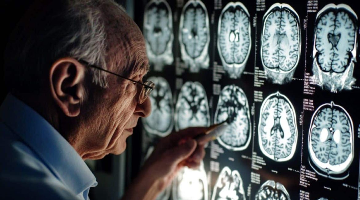
Summary: A new research finds that brain contraction in Alzheimer’s individuals does not following a consistent pattern. Researchers looked through hundreds of brain scans and discovered that individuals displayed various contraction patterns over time.
Those who shrink in particular head regions more quickly were more likely to produce Alzheimer’s. This personal method of brain tracking may lead to custom treatments based on individual mind changes.
Important Information:
- Alzheimer’s people show very varied patterns of brain contraction.
- Poorer memory is correlated to a faster concentration of outcast brain regions.
- Individualized brain maps may help in treatment and disease progression prediction.
Origin: UCL
A new research from researchers at UCL and Radboud University in the Netherlands shows that the brain shrinkage in those who have Alzheimer’s disease does not follow a particular or dress pattern.
Published in , Alzheimer’s &, Dementia, the research is the first to assess unique patterns of brain contraction over time in people with mild storage problems or Alzheimer’s illness against a good benchmark.
Researchers examined mental images for the “fingerprints” of illness and discovered that those with mild memory problems who exhibit more’outlier’ changes more swiftly are more likely to produce Alzheimer’s.
A particular head region that, when adjusted for sex and age, has shrunk more than standard is known as an oddity.
Nonetheless, researchers also found that, despite some coincide, there was no consistent pattern to the way the mind shrank in those who developed Alzheimer’s.  ,
The researchers claim that this new information may lead to the development of more tailored medicines that target the certain range of brain areas affected by a person.
Study writer, Professor Jonathan Schott from UCL Queen Square Institute of Neurology, said:” We know that Alzheimer’s affects everyone differently.
Understanding and analyzing this variation has significant effects on the design and interpretation of clinical trials, as well as, probably, in the future, on the development of personalized treatment plans and treatment strategies.
Alzheimer’s disease is the most common cause of memory, accounting for 60-80 % of the 944, 000 people living with dementia in the , UK.
Past group stage studies have shown that collectively, patients with Alzheimer’s disease have extra mental contraction compared to good controls, and this can be measured using MRI scans.
However, these studies can miss how the pattern of shrinkage varies between individuals, and researchers believe this information could provide valuable information about how an individual’s cognitive performance ( thinking ability/memory recall ) changes over time.
To overcome this, researchers at UCL used a normative modelling1 , approach to gain insights into individual variability between Alzheimer’s patients.
Using data from the Alzheimer’s Disease Neuroimaging Initiative ( ADNI ) they compared 3, 233 MRI brain scans from 1, 181 people with Alzheimer’s disease or mild memory issues to benchmark brain scan data collected from 58, 836 healthy people. The majority of the participants had two or three MRI scans, with the majority taking them at one-year intervals.  ,  ,  ,
Each scan was then processed using special imaging tools, that can assess the structure ( thickness and volume ) of the brain, across 168 different regions.
With this information, researchers were able to create individualized brain maps for each participant, which could be compared to baseline brain maps over time.
The analysis showed that despite most participants starting out with similar-sized brains, different patterns ( progression/regions affected ) of brain shrinkage were seen between individuals over time.
At the start of the study, patients with Alzheimer’s disease typically had 15 to 20 outlier brain regions, which they eventually had after three years. In contrast, patients with mild memory issues typically developed between five and ten outliers and only accumulated two to three more outliers over this time. Importantly, having a higher number of outlier regions was linked to a poorer memory in both groups.
In comparison to those who had mild memory issues, those who went on to develop dementia ( within three years ) accumulated four new outliers annually, whereas in those who continued to have mild issues, the average was less than one new outlier per year.
According to researchers, this new understanding could eventually aid in the prediction of how Alzheimer’s disease will progress based on the initial changes in a person’s brain that have been detected in scans. However, more research is required to identify which brain changes are most likely to predict which symptoms in the future.
Professor James Cole, senior author of the study from UCL Computer Science and UCL Queen Square Institute of Neurology, said:” If we look at which areas of the brain have shrunk most in different people with Alzheimer’s, there’s no single pattern.
The research methodology used in our study allows us to better understand the variation in individual traits in the progression of Alzheimer’s disease. By making these brain maps, which are unique’ fingerprints’ of a patient’s brain health, we can spot if separate brain regions are changing and how rapidly.
Although we’re still a long way from predicting exactly how a person’s disease will progress, this approach should provide insight into how people’s brains can change as symptoms develop and get worse. Hopefully, this is a step towards a more personalised approach to diagnosis and treatment.”
The results revealed that there is a significant overlap in brain shrinkage between healthy people and those with Alzheimer’s and/or mild memory problems for all people who age.
These overlap areas included the hippocampus, amygdala and other parts of the medial temporal lobe known to be critical , for memory, spatial cognition, and emotion.
However, the authors contend that focusing on particular areas may distract from the overall picture by favoring certain ones.
Dr Serena Verdi, first author of the study from UCL Computer Science and UCL Queen Square Institute of Neurology, said:” While it’s true that some regions of the brain, such as the hippocampus, are particularly important in Alzheimer’s disease, we wanted to avoid focusing on specific regions in this study.
Our findings demonstrate that everyone is unique, and that the regions affected by disease in one person may not be the same in the next.
To get away from the notion that” this area is important, this area is n’t,” I believe we need to shift our thinking to a new direction. What counts is the complexity of the larger picture and the variability of people within it.
According to the authors, some of this individual variation may be a result of the fact that many Alzheimer’s patients have multiple causes of cognitive disorders, including vascular or frontotemporal dementia. Genetic and environmental factors, such as brain injuries, alcohol consumption or smoking habits, are also thought to play a part.
About this Alzheimer’s disease research news
Author: Matt Midgley
Source: UCL
Contact: Matt Midgley – UCL
Image: The image is credited to Neuroscience News
Original Research: Open access.
By Jonathan Schott and colleagues,” Personalising Progressive Changes to Brain Structure in Alzheimer’s Disease using Normative Modelling.” Alzheimer’s &, Dementia
Abstract
Using Normative Modelling to Personalize Progressive Changes to Brain Structure in Alzheimer’s Disease
INTRODUCTION
Individual variability in Alzheimer’s disease ( AD ) is captured by neuroanatomical normative modeling. In this study, we used normative modeling to analyze how people’s disease spreads among those with mild cognitive impairment (MCI) and those with AD.
METHODS
Healthy controls ( n , 58k ) were used to create cortical and subcortical normative models. These models were used to calculate regional , z , scores in 3233 T1-weighted magnetic resonance imaging time-series scans from 1181 participants. Regions with , z , scores , <,  , –1.96 were classified as outliers mapped on the brain and summarized by total outlier count (tOC).
RESULTS
TOC increased in AD, MCI-converted AD patients, and there was a correlation between multiple non-imaging markers. Additionally, a higher tOC change rate each year increased the chance of MCI-AD progression. According to brain outlier maps, the hippocampus has the highest rate of change.
DISCUSSION
Regional outlier maps and tOC can be used to track individual patients’ atrophy rates.
