
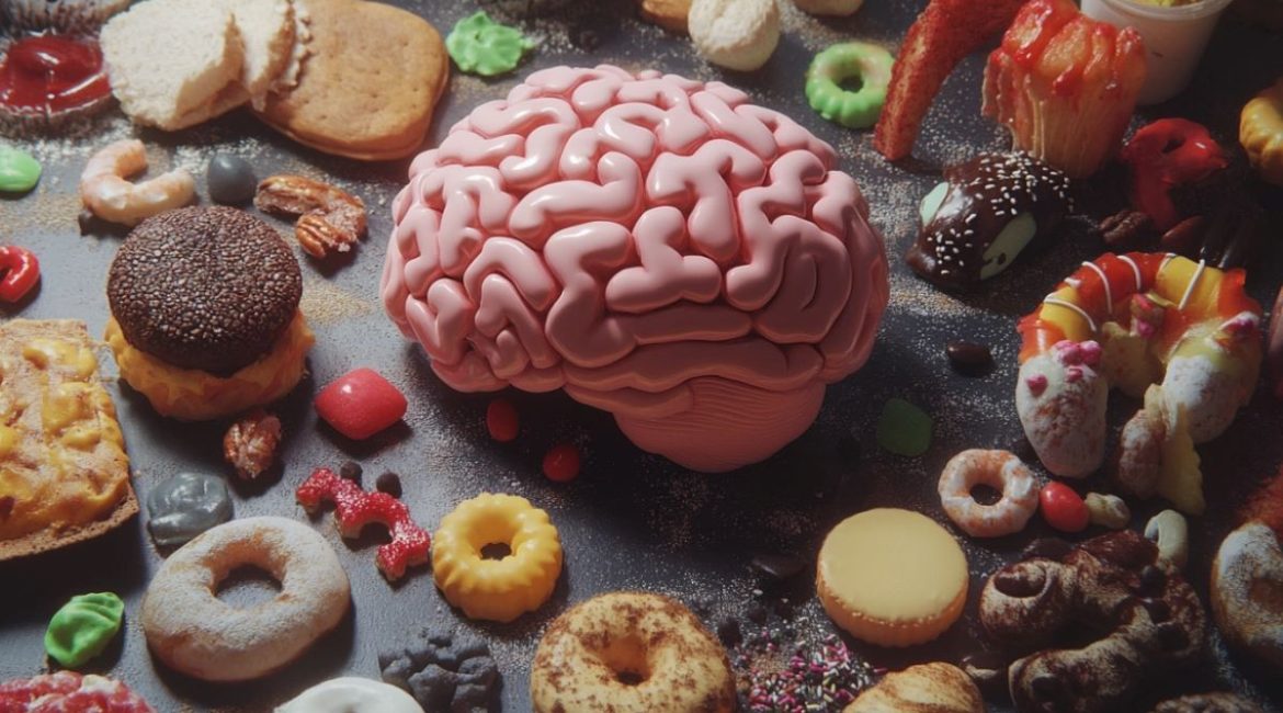
Summary: A recent study demonstrates that changes in brain neurons that control hunger and energy stability are not attributed to one high-fat diet alone. No immediate effects were observed on mice fed a high-fat, low-sugar diet’s brain, suggesting that other nutrients, such as sugar, does have a more significant impact on how brain function is affected.
The research challenges past hypotheses that fat only had a role in affecting energy homeostasis and raising the risk of metabolic diseases. Future research will examine how various macronutrients impact mental function and taste regulation.
Important facts:
- A high-fat eating only did not affect AgRP cells in the brain.
- Another vitamins, such as sugar, perhaps include a more meaningful impact on brain function.
- After 48 hours on the meal, both male and female rabbits showed no increases in nerve connection.
Origin: DZD
A high-fat eating can encourage fat and increase the risk of metabolic disorders, such as diabetes. In animals brains, this leads to measurable alterations in the region of the brain.
However, fat alone does not appear to be responsible for this, as reported by a research team from the German Institute of Human Nutrition Potsdam-Rehbruecke ( DIfE ) and the German Center for Diabetes Research ( DZD ) in the specialist journal , Scientific Reports.
The brain’s links between synapses are continuously evolving. Diet has a major impact on this. A high-fat diet can alter the hypothalamus ‘ ability to disrupt energy balance and raise the risk of metabolic disorders.
Food intake is predominantly regulated within the brain by two types of neurons: AgRP ( Agouti-related peptide ) and POMC ( proopiomelanocortin ) neurons. Both are mostly found in the hypothalamus—or more exactly, in the paraventricular cell, a key area of the hypothalamus—and have same activities. AgRP cells promote meal intake while POMC cells inhibit it.  ,
Large or Instead Sugar?  ,
In mice fed a high-fat dieting, research has demonstrated that AgRP nerve activity in the paraventricular nucleus is decreased. This was most likely due to the high fatty content of the animal nutrition.
However, the meal of the studied animals also contained various nutrients, including honey. Hence, it is impossible to say with certainty which macronutrient causes the cerebral changes.  ,
Scientists from DIfE and DZD examined whether changes in the brain are mainly caused by fat. They fed male and female rabbits a high-fat and low-sugar meal for 48 days.
Because prior research frequently used merely men, it was crucial for the researchers to study both male and female animals. As a result, it was unclear whether the two women respond differentially to a high-fat meal.
Another Nutrients of Greater Significance ,
The evaluation of the animal neurons produced an unforeseen effect: An effect of the high-fat meal was never identified. In either female or male animals, the connection of AgRP cells had no decreased.  ,
This suggests that the earlier observed changes in the brain are not caused by diet alone. The scientists suspect that another nutrient, such as honey, have more serious results on AgRP neurons.
They want to do more research to understand how different macronutrients affect brain neuroanatomical and practical changes.  ,
About this information about science and appetite research
Author: Birgit Niesing
Source: DZD
Contact: Birgit Niesing – DZD
Image: The image is credited to Neuroscience News
Original Research: Start exposure.
Selma Yagoub and colleagues report that “acute increased diet large alone is not sufficient to reduce AgRP forecasts in the paraventricular atom of the brain in mice.” Scientific Information
Abstract
In mice, an acute raised dietary fat level of overweight alone is insufficient to reduce AgRP projections in the paraventricular atom of the brain.
The relationships between cells are constantly changing in the head in response to environmental stimuli. Diet is a significant economic regulator of cerebral activity, and previous research has shown that diet can cause changes in hypothalamic connections when consumed in high amounts of sugar and fat.
We sought to know whether the elevation of dietary fat only contributed to the change in neuroendocrine neuronal connections. Both male and female creatures underwent an examination.
We measured Agouti-related peptide ( AgRP ) neuropeptide and Synaptophysin markers in the paraventricular nucleus of the hypothalamus ( PVH) in response to an acute 48 , h high fat diet challenge.
An experiment using two image examination techniques, as described in earlier studies, did not find an effect on the PVH of male or female animals on AgRP cerebral projections.
These findings point to the possibility that the earlier reported changes in neuroendocrine links may not be caused by diet alone.
Future research should concentrate on understanding how different carbohydrates affect neuroanatomical and useful changes.
