
Summary: Scientists discovered a neuroprotective system in spinal cord injury (SCI) involving microglia, the body’s immune cells, which could provide novel therapeutic goals. The team discovered that microglia form safe wraps around injured axons by using cutting-edge in-vivo scanning, preventing further harm.
This contact, regulated by P2Y12 receptor and potassium channels, sheds light on neuron-glia conversation after injuries. The findings of the study regarding spinal cord safety might eventually lead to novel treatments for SCI and related neurological conditions.
Important Information:
- By wrapping around injured synapses, microglia protect them from future damage.
- Advanced in cellular imaging technologies enabled study of microglia-axon interactions.
- Studies suggest that SCI and additional neurological problems may be treatable.
Origin: HKUST
A neuroprotective mechanism in spinal cord injury (SCI) has been the subject of a collaborative effort between engineers and biologists at the Hong Kong University of Science and Technology ( HKUST ), bringing new insights into potential therapeutic options for millions of patients around the world.
While SCI is a disastrous condition with deep, life-altering consequences, however, a clear cure remains obscure. Lack of reliable in cellular imaging tools that can visualize undisturbed biological processes within the spinal cord has for a long time been a constraint on the study of neuron damage and rejuvenation in SCI.
To overcome this obstacle, an HKUST research group co-led by , Prof. QU Jianan , from the School of Engineering’s Department of Electronic and Computer Engineering (ECE ) and , Prof. LIU Kai , from the School of Science’s Life Science Division, utilized bidirectional microscopy and visual opening technology to enable less aggressive in cellular imaging research.
The crucial role of microglia in post-spinal cable axon injury was revealed during their investigation. The central nervous system’s key immune system cells are microglia. They are known to play important roles in brain development, balance, and neurological problems.
Recent study has underscored the importance of microglial relationships with cells in processes such as neurogenesis, neural plasticity, and degeneration.
Prof. Liu explained,” The existing research, facilitated by cutting-edge in vivo multimodal microscopy and visual opening technology developed in Prof. Qu’s experiment, showcased the ability to comprehend the conversation of glia-nodes in the spinal cord under physiological problems for the first time”.
” Our findings have revealed the neuroprotective capacity of microglia during single spinal cord injury’s acute phase. This unique insight into microglia-axon communication offers promising therapeutic targets for effective treatment strategies”, he said.
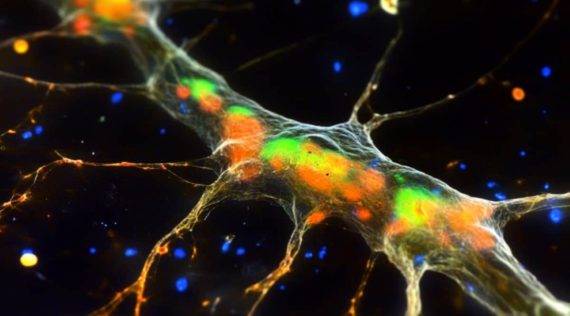
Following an axonal injury, the team demonstrated that microglia exhibit striking neuroprotective wrapping behavior when they come into direct contact with myelinated axons at Ranvier spinal cord nodes.
This protective mechanism, dependent on the function of P2Y12 receptors, highlights a novel form of neuron-glia interaction that prevents acute axonal degeneration from spreading beyond the nodes.
In addition, they found that voltage-gated sodium channels ( NaV ) contribute to the interaction between nodes and glial cells following injury, and that the inhibition of NaV can delay axonal degeneration.
Prof. Qu continued,” The collaborative effort between biologists and engineers has led to the discovery of the remarkable functions of microglia during axonal degeneration and regeneration.” He also emphasized the significance of this discovery.
He added that the study offers hope for the development of novel therapeutic techniques in the future because it not only exposes the intricate mechanisms underlying neuronal diseases.
Our investigation into the causes of various neurological conditions affecting the spinal cord, such as multiple sclerosis, will continue to be fueled by the optical imaging platform developed in this cross-disciplinary study, which are still undetermined. This will be crucial for developing novel, urgently needed treatments, he said.
Their findings have recently been published in the prestigious multidisciplinary journal , Nature Communications. Dr. WU Wanjie ( 2023 ECE PhD graduate ), HE Yingzhu (ECE PhD student ), and CHEN Yujun ( Life Science PhD student ) are the co-first authors, while Prof. Qu and Prof. Liu are the corresponding authors.
About this news from neurology and SCI research
Author: Victor Lee
Source: HKUST
Contact: Victor Lee – HKUST
Image: The image is credited to Neuroscience News
Original Research: Open access.
” In vivo imaging of mouse spinal cord reveals that microglia prevent degeneration of injured axons,” by QU Jianan and al. Nature Communications
Abstract
In vivo imaging of a mouse spinal cord revealed that microglia prevent the injured axons from deteriorating.
Through their interaction with neurons, microglia, the primary immune cells in the central nervous system, play a crucial role in controlling neuronal function and fate. Despite extensive research, the specific functions and mechanisms of microglia-neuron interactions remain incompletely understood.
In this study, we show that microglia in the spinal cord of mice have direct contact with myelinated axons at Nodes of Ranvier. The neuronal contact is made in a random scanning pattern.
Axonal injury causes microglia to quickly transform into a strong wrapping form, preventing acute axonal degeneration from extending beyond the nodes.
The microglial P2Y12 receptors ‘ ability to wrap themselves depends on their function, which may be activated by ATP released through axonal volume-activated anion channels at the nodes.
Additionally, voltage-gated sodium channels ( NaV ) and two-pore-domain potassium ( K2P ) channels contribute to the interaction between nodes and glial cells following injury, and inhibition of NaV delays axonal degeneration.
Through in vivo imaging, our findings reveal a neuroprotective role of microglia during the acute phase of single spinal cord axon injury, achieved through neuron-glia interaction.
