Summary: New research has revealed a straightforward neural circuits that links the jaw activities necessary for eating. Experts identified a three-neuron road involving the hormone leptin and BDNF cells, which controls the jaw’s engine performance.
When these cells were activated, animals stopped eating, when inhibited, they made chewing movements yet without foods. The findings provide novel insights into how poverty and motor control are related, suggesting that feeding habits does resemble a reaction.
Important Information:
- A three-neuron loop connects appetite signals to chewing activities.
- Inhibiting BDNF cells causes compulsive sucking, yet without food.
- Feeding conduct may work like a reaction, driven by this simple neurological circuit.
Origin: Rockefeller University
Speaking, singing, breathing, laughing, yelling, yawning, chewing—we employ our mouths for several reasons. Every movement necessitates a difficult coordination of muscles whose activity is controlled by brain cells.
However, it turns out that the neural circuits that controls eating, the most crucial component of survival, is amazingly straightforward, as recent researchers from Rockefeller University have demonstrated in a new document in Nature. Christin Kosse and other experts from the , Laboratory of Molecular Genetics, headed by Jeffrey M. Friedman, have identified a three-neuron loop that connects a hunger-signaling testosterone to the neck movements of munching.
A swarm of cells in a particular area of the brain, which has long been associated with obesity, serves as the middleman between these two.
Strangely, inhibiting these so-called Hippocampal neurons causes the jaw to make chewing motions even when there is no food or other sensory input that may show it was time to eat. It also causes animals to eat more food. And by stimulating them, one can reduce food intake and stop chewing, effectively reducing poverty.
The straightforward design of this loop suggests that the eating desire may be more similar to a reaction than previously thought and may offer new insights into how feeding is initiated.  ,
According to research first publisher Christin Kosse, a study connect in the laboratory, “it’s surprising that these cells are so keyed to motor control.” ” We did n’t expect that limiting physical jaw motion could act as a kind of appetite suppressant” . ,
More than a experience?
Not only thirst, but many other factors are involved in the drive to eat. We also eat for joy, area, ceremony, and behavior, and smell, taste, and emotions may effect whether we eat too. Eating can also be regulated by the unconscious desire to consume more or less in humans.
The causes of obesity are equally complex, the result of a dynamic interplay of diet, environment, and genes. For example, mutations in several genes, including leptin, the hunger-suppressing hormone, and brain-derived neurotrophic factor ( BDNF), lead to gross overeating, metabolic changes, and extreme obesity, suggesting that both factors normally suppress appetite.
When Friedman’s team began this study, they sought to pinpoint the location of the BDNF neurons that curtail overeating. That’s eluded scientists for years, because BDNF neurons, which are also primary regulators of neuronal development, differentiation, and survival, are widespread in the brain.  ,
In the current study, they homed in on the ventromedial hypothalamus (VMH), a deep-brain region linked to glucose regulation and appetite. It is well known that damage to the VMH can cause overeating and eventually obesity in both animals and people, just as mutated BDNF proteins do. Perhaps the VMH regulated feeding behavior.
They hoped to discover the neural circuit responsible for the conversion of sensory signals into jaw motions by documenting BDNF’s impact on eating behavior.
After that, they discovered that BDNF neurons in the VMH, but not elsewhere, are activated when animals become obese, suggesting that they are activated when they gain weight to enslave themselves. Thus when these neurons are missing, or there is a mutation in BDNF, animals become obese.
Chewing without food
The researchers then used optogenetics to either express or inhibit the BDNF neurons in the mice’s ventromedial hypothalamus during a series of experiments. When the neurons were activated, the mice completely stopped feeding, even when they were known to be hungry.
Silencing them did the opposite: They started eating, eating, eating, and eating, eating, eating, wolfing down nearly 1200 % more food than they normally would in a short amount of time.
” When we saw these results, we initially thought that perhaps BDNF neurons encode valence”, Kosse says.
” We wondered if the mice were experiencing the negative sensation of hunger or perhaps the positive sensation of eating delicious food,” said the researchers.
However, later tests disproved that hypothesis. Regardless of the food consumed by the mice, such as their standard chow or food that is fat and sugary, like the chocolate mousse cake-like diet that is consumed by mice, the researchers discovered that activating the BDNF neurons prevented food intake.
And because everyone who ca n’t skip dessert can attest that they were already well-fed, they also provided high-palatable food to mice. The animals chowed down until the researchers blocked the BDNF neurons, at which point they abruptly stopped eating.  ,
This was initially a perplexing finding because previous studies have suggested that this “hedonic” drive to eat for pleasure is quite different from the hunger drive, which is an attempt to suppress the negative feeling, or negative valence, associated with eating, Kosse notes.
” We demonstrated that both drives can be suppressed by activating BDNF neurons.”
Even when there was no food nearby, the mice made similar chewing motions with their jaws that were caused by BDNF inhibition. The mice gnawed on anything that was around them, including the wires monitoring their neural activity, as well as the metal spout of a water feeder, a block of wood, and even the wires monitoring their neural activity.
The circuit
But how does the body’s desire or need for food relate to this motor-control switch?
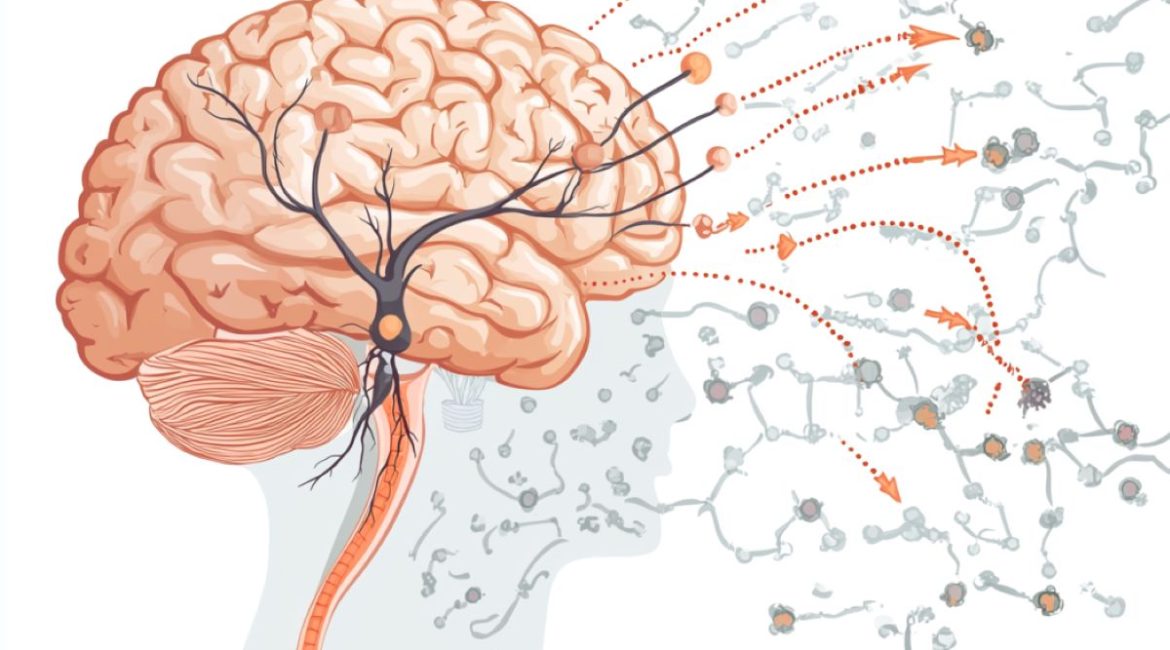
The researchers discovered that BDNF neurons are the linchpin of a three-part neural circuit that links hormonal signals that regulate appetite to the movements required to consume it by mapping the inputs and outputs of the BDNF neurons.
At one end of the circuit are special neurons in the arcuate nucleus ( Arc ) region of the hypothalamus that sense hunger signals like the leptin hormone that fat cells produce. A high leptin level indicates that the energy tank is full, while a low leptin level indicates that food is in order. Animals without leptin become obese.
The BDNF neurons direct the Arc neurons ‘ signals to a brainstem center called Me5 that controls the movement of jaw muscles by projecting to the ventromedial hypothalamus.
” Other studies have shown that when you kill Me5 neurons in mice during development, the animals will starve because they’re unable to chew solid foods”, says Kosse. ” So it makes sense that we can see jaw movements when we manipulate the BDNF neurons that are protruding there.”
It also explains why damage in the VMH causes obesity, Friedman says. The findings unify the known mutations that cause obesity into a relatively coherent circuit and support the hypothesis that the obesity linked to these lesions is caused by a loss of these BDNF neurons.
He continues,” The findings suggest something more about the relationship between sensation and behavior.” According to Friedman,” the architecture of the feeding circuit is not very different from the architecture of a reflex.”
” That’s surprising, because eating is a complex behavior—one in which many factors influence whether you’ll initiate the behavior, but none of them guarantee it. On the other hand, a reflex is simple: a defined stimulus and an invariant response.
The line between behavior and reflex is probably more blurred than we thought, according to this paper. We make the hypothesis that the neurons in this circuit are the targets of other brain neurons that generate other signals that regulate appetite.
This hypothesis is consistent with the work of early 20th , century neurophysiologist , Charles Sherrington, who pointed out that while cough is regulated by a typical reflex, it can be modulated by conscious factors, such as the desire to suppress it in a crowded theater.
Kosse adds”, Because feeding is so essential to basic survival, this circuit regulating food intake may be ancient. It might have served as a substrate for more intricate processing as the brain developed.
Future research projects the brainstem area known as Me5 with the intention that the researchers” jaw’s motor controls could serve as a model for understanding other behaviors, including gnawing on a pencil eraser or strands of one’s hair.
She claims that by looking at these Me5 premotor neurons, we might be able to determine whether other centers are present in the area and have an impact on other innate behaviors, such as BDNF neurons ‘ ability to control eating. Are there any stress-activated or other neurons there that also protrude into it?
About this news from neuroscience research
Author: Katherine Fenz
Source: Rockefeller University
Contact: Katherine Fenz – Rockefeller University
Image: The image is credited to Neuroscience News
Original Research: Open access.
A subcortical feeding circuit by Christin Kosse and al. connects an interoceptive node to jaw motion. Nature
Abstract
An interoceptive node and jaw movement are connected through a subcortical feeding circuit.
The brain processes a large range of stimuli, allowing for the selection of appropriate behavioral responses, but the neural networks that link interoceptive inputs to feeding outputs are poorly understood.
In the ventromedial hypothalamus (VMH), brain-derived neurotrophic factor ( BDNF)-expressing neurons directly connect interoceptive inputs to motor centers, which are in charge of controlling food consumption and jaw movements.
VMHBDNF , neuron inhibition causes the jaw to be gated to produce more food by promoting motor-sequence feeding patterns. When food is unavailable, VMHBDNF , inhibition elicits consummatory behaviours directed at inanimate objects such as wooden blocks, and inhibition of perimesencephalic trigeminal area ( pMe5 ) projections evokes rhythmic jaw movements.
These neurons are more active when food is in proximity but not consumed, and their activity is decreased when eating. In obese animals and after receiving leptin, activity is also increased. VMHBDNF , neurons receive monosynaptic inputs from both agouti-related peptide ( AgRP ) and proopiomelanocortin neurons in the arcuate nucleus ( Arc ), and constitutive VMHBDNF , activation blocks the orexigenic effect of AgRP activation.
These data point to an Arc- VMHBDNF– pMe5 circuit that senses an animal’s energy state and regulates consummatory behaviors in a state-dependent manner.
