Summary: Experts have uncovered a process that may cause ALS’s earliest phases, identifying proteins that mislocalize, causing nerve degradation. By preventing important Sind proteins from leaving their protecting cellular regions and maintaining nerve function by targeting the RNA-binding proteins SmD1, scientists were able to do this.
The findings offer prospective new ways to treat the disease, including ALS therapies that stop progression before considerable neurodegeneration occurs.
Important Facts:
- ALS growth does started when the amino CHMP7 mislocalizes, initiating nerve damage.
- Restoring SmD1 functionality may reduce CHMP7 mislocalization and guard neurons.
- By stabilizing nerve dignity, risdiplam and other medications like risdiplam may slow the onset of ALS.
Origin: UCSD
Approximately 5, 000 people in the U. S. develop amyotrophic lateral sclerosis ( ALS ) each year. On average, they survive for just two to five years after being diagnosed, according to the Centers for Disease Control and Prevention.
Neurological disease, which is rapidly progressing, causes dementia, respiratory failure, and muscle weakness.
Little is known about the initial causes of the disease’s damaging impact on engine neurons.
Researchers from the University of California San Diego and their associates now report that they have discovered a crucial mechanism that causes aging in the disease’s early stages.
The results could lead to the development of treatments to stop or stop the progression of ALS before it has already caused significant harm.
The study was published on October 31, 2024 in , Neuron.
TDP-43, a protein that controls the performance of motor neurons, is typically found in the nucleus of the cell. Research have shown that when TDP-43 rather accumulates in the nucleus, outside of the centre, it is a telltale sign of ALS. How the proteins ends up in the wrong place, leading to neuronal degradation, has stumped experts until now.
” By the time you see a person with ALS and you see the TDP-43 proteins aggregated in the nucleus, it’s like the crash site with all the cars crashed now, but that’s not the initiating event”, said related author Gene Yeo, Ph. Dr., director of the Center for RNA Technologies and Therapeutics and the Innovation Center at the Sanford Stem Cell Institute, and teacher of the San Diego School of Medicine’s Department of Cellular and Molecular Medicine.
Tracing the events leading up to the “accident”, Yeo explains that another proteins, called CHMP7 — usually found in the cytoplasm — builds up in the cell otherwise, setting off a cascade of events that eventually lead to engine nerve degradation. What, then, is the beginning of CHMP7 accumulating in the cell?
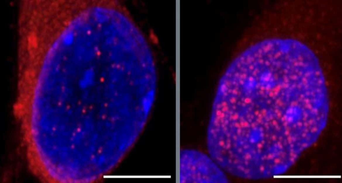
Harrison and his team screened for RNA-binding molecules that may affect CHMP7 build-up in the cell. This yielded 55 enzymes, 23 of which had a possible connection to ALS pathogenesis. A rise in CHMP7 in the cell was caused by inhibiting the production of several of these protein.
The most significant increase in nuclear formation was caused by the discovery that one of the following, an RNA-splicing associated proteins called SmD1, which had not originally been known to affect CHMP7 rates, had been depleted by additional experiments with motor neurons created from ALS-patient-derived induced pluripotent stem cells.
Nucleoporins, which Yeo refers to as little portals in the barrier that control the movement of protein and RNA between the two mobile areas, are damaged by a build-up of CHMP7 in the cell. TDP-43 can leave the nucleus and build up in the cytoplasm thanks to malfunctioning nucleoporins. The protein no more manages the gene expression processes necessary for cells to function.
However, when the researchers boosted SmD1 appearance in cells, CHMP7 was restored to its normal place in the nucleus, leaving nucleopores alive, allowing TDP-43 to stay in the cell, so sparing the engine neurons from degradation.
Norah Al-Azzam, the first author of the study, a neurosciences student in the Yeo lab who later earned her Ph. D., claimed that “you can actually fix the localization of this CHMP7 protein and all of the downstream effects.” D. in spring 2024.
What’s more, the SmD1 protein is part of SMN, a multiprotein complex. Spinal muscular atrophy, a neurodegenerative disorder, is a result of SNM dysfunction.
” We’re intrigued because there are actually therapeutics for spinal muscular atrophy”, Yeo said. Risdiplam, a small molecule substance, “enables the splicing and expression of SMN2, a gene closely related to the SMN1 gene that malfunctions in ALS,” says one of them.
This suggests that lowering SMN levels with risdiplam might help delay the disease’s onset until its most advanced stage.
” It’s not like all the neurons die at once”, said Yeo. ” Some neurons die first, and then there is spread across other neurons. We might start treating the patient so that the rest of the neurons do n’t crash and hope that you stop the ALS progression as soon as you start to show symptoms.
The SMN complex, according to the researchers, may be responsible for the development of ALS, but more research is required. The next step will be to raise money to continue the research in ALS-specific animal models and genetic models, and to evaluate the potency of risdiplam or other medications for short-circuiting ALS.
Additional co-authors on the study include: Jenny H. To, Vaishali Gautam, Dylan C. Lam, Chloe B. Nguyen, Jack T. Naritomi, Assael A. Madrigal, Benjamin Lee, Anthony Avina, Orel Mizrahi, Jasmine R. Mueller, Willard Ford, Anthony Q. Vu, Steven M. Blue, Yashwin L. Madakamutil, Uri Manor, Cara R. Schiavon and Elena Rebollo, all at UC San Diego, Wenhao Jin at Sanford Laboratories for Innovative Medicines, Lena A. At Johns Hopkins University School of Medicine, Straet and Marko Jovanovic, and Jeffrey D. Rothstein and Alyssa N. Coyne.
Funding: The study was funded, in part, by the National Institutes of Health ( grants R01HG004659, U24 HG009889, R35GM128802, R01AG071869 and R01HG012216 ), the National Science Foundation (MCB-2224211 ), and the Chan-Zuckerberg Initiative.
About this news from ALS research
Author: Susanne Bard
Source: UCSD
Contact: Susanne Bard – UCSD
Image: The image is credited to UCSD
Original Research: Open access.
” Inhibition of RNA splicing triggers CHMP7 nuclear entry, impacting TDP-43 function and leading to the onset of ALS cellular phenotypes” by Gene Yeo et al. Neuron
Abstract
Inhibition of RNA splicing triggers CHMP7 nuclear entry, impacting TDP-43 function and leading to the onset of ALS cellular phenotypes
Certain nucleoporins in neurons have been reduced, according to Amyotrophic lateral sclerosis ( ALS ). A protein involved in nuclear pore surveillance, charged multivesicular body protein 7 ( CHMP7 ), has been identified as a major contributor to the destruction of nuclear pores and the disintegration of transport.
Using CRISPR-based microRaft, followed by gRNA identification ( CRaft-ID), we discovered 55 RNA-binding proteins ( RBPs ) that influence CHMP7 localization, including SmD1, a survival of motor neuron ( SMN) complex component.
Immunoprecipitation-mass spectrometry ( IP-MS ) and enhanced crosslinking and immunoprecipitation ( CLIP ) analyses revealed CHMP7’s interactions with SmD1, small nuclear RNAs, and splicing factor mRNAs in motor neurons ( MNs ).
ALS induced pluripotent stem cell (iPSC ) -MNs show reduced SmD1 expression, and inhibiting SmD1/SMN complex increased CHMP7 nuclear localization.
Crucially, overexpressing SmD1 in ALS iPSC-MNs restored CHMP7’s cytoplasmic localization and corrected STMN2 splicing. Our findings support the role of SMN complex dysregulation in early ALS pathogenesis.
